Download
1 / 40
460 likes | 1.65k Views
Allergic Conjunctivitis. Treatment: Eliminate causative factor Cool compresses several times daily Pharmacologic treatment Mild – artificial tears qid, mast cell stabilizers Moderate – vasoconstrictor/antihistamine (naphazoline) qid, systemic antihistamines

E N D
Allergic Conjunctivitis Treatment: • Eliminate causative factor • Cool compresses several times daily • Pharmacologic treatment • Mild – artificial tears qid, mast cell stabilizers • Moderate – vasoconstrictor/antihistamine (naphazoline) qid, systemic antihistamines • Severe – topical antihistamine such as olapatidine, mild topical steroid qid if keratopathy (would let ophthalmologist manage topical steroids)
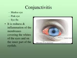
Viral Conjunctivitis • AKA “pink eye” • Symptoms – watery discharge, red swollen eyelids, often bilateral • Signs – conjunctival injection, edema, and follicles, pre-auricular node common • Often preceded by upper respiratory tract infection or contact with someone with red eye
Viral Conjunctivitis • Treatment – palliative only • Cold compresses • Artificial tears • Erythromycin • Very contagious • Don’t share towels, etc. • No school
Bacterial Conjunctivitis • Symptoms – irritation, yellow/green discharge, mattering of eyelashes • Signs – conjunctival papillae, injection, and chemosis; subconjunctival hemorrhage possible in severe cases • Treatment – topical antibiotics • Trimethoprim/polymyxin B or tobramycin qid x 4-7 days. Cover S. pneumoniae, H. influenzae, Moraxella, Staphylococcus. • Ciprofloxacin in contact lens patients? • Gram stain and culture if copious discharge or suspicion of STI? Neisseria gonorrheae is a possible cause. IV antibiotics if GC.
Corneal Abrasion • Treatment – topical antibiotic • Ointment vs. solution • Erythromycin ointment • Topical cycloplegic • Cyclopentolate 1% • Oral analgesics • Discontinue any contact lens wear • Do not pressure patch
Corneal Foreign Body • Treatment • Give topical anesthetic prior to removal • Cyclopentolate 1% • Erythromycin ointment • Needs orbital X-ray if high velocity suspected – rule out intravitreal foreign body • Patient education concerning safety glasses
Keratitis • Corneal ulcer due to infection or systemic diseases • Bacterial, fungal or treponemal diseases, connective tissue disorder • Cornea will appear opaque, WBC seen in anterior space • Herpes keratitis presents with characteristic branching dendrite pattern on corneal exam
Iritis/Uveitis • Anterior chamber inflammation secondary to injury, infection, collagen vascular disorder • Treatment • Cycloplegic (cyclopentolate 1%) • Ophthalmologist may consider topical steroid • Needs dilated retinal examination • Must rule out corneal abrasion
Acute Angle Closure Glaucoma • Sudden complete closure of angle, resulting in intraocular pressures of 50-100 mmHg • Patient complaints of severe pain, nausea, vomiting and decreased visual acuity • Eye is red, cornea is steamy and edematous, pupil is fixed in mid dilation, i.e. not a subtle presentation • Treat as emergency – hospitalize, acetazolamide, mannitol, surgery
Primary Open Angle Glaucoma • POAG is asymptomatic, even at pressures up to 40 mmHg. There is no loss of visual acuity and patients may not notice loss of peripheral vision when it has progressed to central "tunnel vision”. • Intraocular pressure measurement (IOP) alone is not a satisfactory for diagnosis. One-third to one-half of patients with visual field defects due to glaucoma have an IOP <21 mmHg, while many with pressures over 21 mm HG have no optic nerve damage. • The fundus examination is part of the routine medical physical examination. Presence of cupping, a cup that is greater than 50 percent of the vertical disc diameter, is a useful threshold for suspicion of glaucoma.
THE INTERNIST AND THE EYE Clinical Findings and Ophthalmic Manifestations of Systemic Diseases
Eyelids, Conjunctiva, and Cornea • Band keratopathy • Deposition of calcium in cornea • Appears at 3 and 9 o’clock to gross eye examination • Goes beyond limbus • Horizontal grayish bands where the eyelids close can be seen on slit-lamp exam
Clinical Findings Anterior Chamber
Clinical Findings Iris and Pupils
Clinical Findings Fundus
Fundus • Hypertensive retinopathy • Keith-Wagner-Barker stages • Stage I – moderate arteriolar narrowing • Stage II – arteriovenous crossing changes and arterioles narrowing to one-half normal size • Stage III – above plus cotton wool spots (ischemic infarcts), hemorrhages may also be present • Stage IV – above plus papilledema