Download
1 / 34
391 likes | 1.58k Views
Muscular System. Motor Unit: There is a decrease in motor units with age. The number of ... Active VitD assists in actively transporting Ca++ through the system ...

E N D
Slide 1:Musculoskeletal Changes Associated with Aging
Slide 2:Muscular System
Motor Unit: There is a decrease in motor units with age The number of motorneurons dec with age Muscle fibers also dec with age Slow twitch Type I- there is a higher percentage of this type Fast Twitch Type II- are selectively lost to a greater degree
Slide 3:Morphological and Functional Age related Changes
Muscle mass: From age 40 to age 80 there is a 30-40% decrease in muscle mass. It is obvious by 60. Antropometrically, there is a decrease in lean muscle mass and an increase in fat Muscle fiber size decreases with inactivity in both slow and fast twitch. Since there is no decrease in enzymes related to energy metabolism in skeletal muscle, aerobic and anaerobic metabolism does Not decline 2ary to aging
Slide 4:Aging: Joint mobility
There is a decrease in extremes of joint ROM even without pathology PROM decreases with age Jt. Flexibility is inversely related to age. Women lose ROM at a slower rate than men. UE joints remain more flexible then LE PCT stiffness contributes to dec. ROM due to changes in collagen structure, dec. physical activity
Slide 5: Arthrokinesiological Changes
There is decreased angular velocity and displacement. Angular velocity Parallels decreased overall physical activity Other contributing factors are decreased m strength, meds, fear of falling
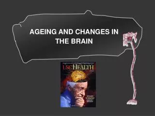
Slide 6:Sensorimotor changes
Dec. reaction times Inc. rate of loss of brain cells Altered neurotransmitter production Dec. perception of vibration, temp, touch, proprioception and pressure When these factors are put together, there is a dec. rate or magnitude of force generated by muscle
Slide 7:Age Related joint mechanics Changes
Arthrokinematics are affected by increased PCT stiffness when it interferes with natural translation of the joint. Gains can be made by stretching Sometimes these reductions produce enhanced safety
Slide 8:Effects of exercise training on muscle performance
When engaged in programs of 6-25 weeks of sufficient intensity, elders have shown significant increases in strength. 70-80% of 1 REM is considered high intensity. Lower intensities will also improve strength. Resistance exercises are better than walk/jog to increase general ex tolerance. There is a poor correlation between strengthening and function
Slide 9:Skeletal Changes
Bone mineral density declines with age After age 60, bone density declines at a rate of about 1%/year for both sexes. By age 90, women lose 30% cortical bone, and men lose 20%
Slide 10:About Fractures
About � of all hospital admissions for fractures is secondary to hip fractures. ~90% of hip fractures result from falls 1/3 of all females > 65 will have a vertebral fracture The forearm (radius) is the third most common fracture
Slide 11:Factors contributing to bone loss
Dec Ca++ in diet Dec Ca++ absorption Hormonal changes Lack of exercise Gender Caffeine Genetics alcohol cigarettes
Slide 12:Effects of Exercise on the Skeletal system
Dec. bone loss. However ex must be done for 9 months to 1 year to increase bone density. It can be strengthening or aerobic
Slide 13:Bone density
Low bone density is defined as 1.0gm/cm2. This is also considered the fracture threshold. Ca++ supplementation will not be effective in reducing the loss occurring during the first 5 post menopausal years. Bed rest has a more profound effect on loss of bone then the above
Slide 14:CA++ role in bodily functions
M contraction N conduction Cell membrane maintenance Blood clotting
Slide 15:Regulating Ca++
PTH- prevents hypocalcaemia. PTH makes sure the kidney gets 9 gms of Ca++/day for reabsorption at the nephron. Osteoclastic cells are sensitive to PTH. In the GI tract, PTH assists with Ca++ absorption Vitamin D: Active Vitamin D is a hormone that is converted first in the liver then the kidneys. Active VitD assists in actively transporting Ca++ through the system
Slide 16:Regulating Ca++ con�t
Calcitonin, excreted by the thyroid, assists in depositing Ca++ in the bone Estrogen until menopause, protects females from osteoporosis RDA Ca++ young adults 750-1000mg., premenopausal 1000mg, preg/postmeno1500mg. Vitamin D 400IU hopefully � from sun, 1/2 from diet
Slide 17:Remodeling
Each day 15% of the skeleton is being remodeled. The osteoclast goes into the bone and excavates, this takes ~1 month, then reintroduces Ca++ back to the circulation. The osteoblast however, needs three months to fill in that hole. Therefore you need enough Ca++ and have osteogenic stimulus provided by exercise to sustain bone mineral density Peak bone mass occurs at 35 years. Highest amt. Of Ca++
Slide 18:Posture
Normally, external forces created by the body are favorable for energy conservation. In elders, when mobility becomes limited, forces acting on the joints produced by gravity are no longer efficacious .
Slide 19:What�s different about the posture of an elder?
Increased thoracic kyphosis Decreased lumbar lordosis Posterior pelvic tilt Forward head, rounded shoulders Flexed hips and knees Tight gastrocs/soleus
Slide 20:REEDCO Posture Scoring
Used to assess static posture. Score from 100 (perfect) to 0 (poor) Allows scoring over 4 occasions. Provides a venue for quantifiable documentation of improvement.
Slide 21:Osteoporosis
Preventable Fantastic venue for promotion and wellness throughout the lifespan Children should be educated regarding intake, avoidance of risk factors, exercise Young women should be made aware during childbearing age of the successful management and prevention Peri/postmenopausal women
Slide 22:What is available for prevention of Osteoporosis today?
HRT Nutritional Interventions Physical Activity Bone enhancing: Need a mechanical load Must realize they have to continue
Slide 23:Osteoporosis Education
Optimal skeletal alignment Avoidance of postures and positions putting a bone at fracture risk Avoid spinal flexion exercises Generally stretching the anterior structures
Slide 24:Examination
Take an exercise inventory Type Frequency Duration Intensity
Slide 25:Tests and Measures
Special Attention to Posture Balance: Functional Reach Gait Scapula m strength Body mechanics 6 minute walk test
Slide 26:Acute Fracture Management
Teach posture Body mechanics Make sure pt. Is up at least 10 minutes out of every waking hour and gradually increase Walking is important, rolling support if necessary Sit in a firm but comfortable chair
Slide 27:Exercises During Recovery
Isometric Trunk Extension Chin Tuck �V� exercise �W� exercise Money exercise
Slide 28:Commonly used hip fixation devices
Slide 29:Hip Fixes
Slide 30:Risk Factors for Hip Fractures
Female White Low weight Physical inactivity Cognitive impairment Old age Pyschotropic meds Estrogen deficiency High levels ETOH Caffeine Reduced m strength LE Impairment of postural control Neurological cond. CVA,PD
Slide 31:Hip Fracture Sites
Most common are intertrochanteric and femoral neck Intertrochanteric usually pt. Has osteoporosis Femoral neck � circulation is a concern Subtrochanteric 10% of all fx. % cm. Distal to lesser trocanter
Slide 32:Outcomes?Rehab Hip FX
Functional independence is achieved more in: 1. Pts. < 85 years 2.Had no post op complications 3.PT BID in acute care 4.I in bed mob., transfers, amb with walker
Slide 33:One year post op
92% were amb if they were amb before Only 41% regained prefracture status
Slide 34:Case Studies
What practice pattern? Which T&M What additional considerations? What are the goals of your interventions and treatment options?