Download

1 / 15
250 likes | 3.01k Views
Embryology of the Human Ear. Ashley Balaker, M.D. March 17, 2010. Outline. Inner Ear Middle Ear External Ear. Inner Ear. Ectoderm = membranous labyrinth Mesoderm = bony labyrinth 3rd week Surface ectoderm = otic placode Invaginates --> otic pit 4th week

E N D
Embryology of the Human Ear Ashley Balaker, M.D. March 17, 2010
Outline • Inner Ear • Middle Ear • External Ear
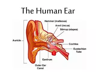
Inner Ear • Ectoderm = membranous labyrinth • Mesoderm = bony labyrinth • 3rd week • Surface ectoderm = otic placode • Invaginates --> otic pit • 4th week • Pit edges fuse to become otocyst
Inner Ear • Dorsal utricular portion, ventral saccular portion • Utricular portion • 3 diverticula for semicircular canals • Saccular portion • Tubular diverticulum (cochlear duct) grows in spiral fashion to become membranous cochlea • The organ of Corti differentiates from cells along the wall of the cochlear duct.
Inner Ear • 6th week • neuroectoderm --> spinal and vestibular ganglia and corresponding sensory nerves • Mesoderm around otocyst soon forms a cartilaginous otic capsule. • Ossifies by 25 weeks
Inner Ear • Vacuoles containing the perilymph develop within the otic capsule. • The vacuoles enlarge and unite to form the perilymphatic space • Divides into the scala tympani and the scala vestibuli. • The cartilaginous otic capsule ossifies to form the bonylabyrinth of the inner ear (mesoderm).
Middle Ear • 1st pharyngeal pouch endoderm • Lining of middle ear (tympanic cavity) • Connection to pharynx elongates and forms eustachian tube
Middle Ear • 1st and 2nd pharyngeal arch cartilage (mesoderm) --> ossicles • 1st (Meckel’s): epitypanum ossicles • Head of malleus, body and short process of incus • 2nd (Reichert’s): mesotympanum ossicles • Long process malleus, long process incus, stapes superstructure • Stapes footplate: otic capsule
Middle Ear • Ossicles full sized by 15 weeks • Ossify by 25 weeks
External Ear • EAC develops from the surface ectoderm that covers the dorsal end of the first pharyngeal grove. • A solid epithelial plate meatal plug develops at the bottom of the funnel-shaped pharyngeal groove.
External Ear • TM • Inner layer endoderm • Middle layer mesoderm • Outer layer ectoderm
Auricle • 6 Hillocks of His (mesoderm) • 1st pharyngeal arch • 1: Tragus • 2: Helical crus • 3: Helix • 2nd pharyngeal arch • 4: antihelix • 5: antihelix • 6: antitragus