Download
1 / 2
20 likes | 501 Views
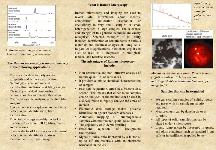
Spectrum of cocaine taken through a polyethylene bag. What is Raman Microscopy

E N D
Spectrum of cocaine taken through a polyethylene bag What is Raman Microscopy Raman microscopy and imaging are used to reveal vital information about identity, composition, molecular orientation or crystallinity in very small samples or small heterogeneities in large samples. The relevance and strength of this generic technique are widely recognised. Selected examples of its utility include: identification of contaminants in various materials and chemical analysis of living cells. In parallel to applications in biochemistry, it can also be used as a diagnostic in biological, medical and forensic sciences. A Raman spectrum gives a unique chemical signature of a specimen • The advantages of Raman microscopy include: • Non-destructive and non-intrusive analysis of minute quantities of substances • It provides a unique chemical characterisation of samples • Fast data acquisition, often in a fraction of a second. This means that either more samples can be analysed or the method can be used in a survey mode to rapidly analyse the areas of interest • Electronic data storage makes possible identification against standard databases • Automatic mapping of inhomogeneous samples with micrometer spatial resolution • The capability of in-depth profiling • Excellent rejection of background fluorescence • Signal to noise ratio improved by a factor of up to 106 (in materials with an electronic resonance in the UV) • The Raman microscopy is used extensively in the following applications: • Pharmaceuticals – for polymorphs, excipients and actives identification • Gemology – gem and mineral identification, inclusion and filing analysis • Chemistry – catalyst composition, ceramics, oxides and many other areas • Corrosion – oxide analysis, protective film analysis • Forensic science – explosive and narcotics detection and identification, fibre identification • Protective coatings – quality control of diamond-like-carbon (DLC) films, paints and adhesives • Semiconductors/Electronics – contaminant detection and identification, stress measurements, surface damage Mixture of cocaine and sugar. Raman image (right) reveals particles of cocaine indistinguishable in a standard microscope image (left). • Samples that can be examined • We can examine samples of solids, liquids and gases with no sample preparation required. • Measurements can be done in air or in solution. • All types of solid samples that can be placed under a microscope • Liquid samples:can be measured in quartz and glass containers such as standard 1 cm cells or in capillaries (supplied by us)
Do you need chemical characterisation at a microscopic level? • Macquarie University • Raman Microscope • Technical specifications • Excitation wavelengths of 325 nm and 406 nm are available • Spectral resolution – 3 cm-1 • Spectral range (Raman shift) 400-4000cm-1 • Spatial resolution – 2 micrometres (x 40 objective), 1 micrometres (x 100 objective) • Depth of field 2 micrometres ( x 100 objective) • Measurements are done in 180 degree backscattering configuration • Polarisation accessories are available • Extended scan option gives UV Raman and fluorescence spectra at the same point and time • Resonance enhanced signals • Reduced fluorescence effects • Measurements can be made at all temperatures between 77K and 600K Competitive pricing The facilities and expertise of the Optical Characterisation Facility are available to the general community. For external customers that require Facility staff to run the instrument for them the charge is $99 per hour, for those who are able to operate the system the charge is $66 per hour. A minimum charge of 30 minutes applies. Lower rates apply to internal users. All prices are inclusive of GST and a tax invoice will be issued. Data interpretation services are charged separately. For more information contact Associate Professor Ewa M. Goldys Division of Information and Communication Sciences Macquarie University, North Ryde 2109 NSW, Australia email: goldys@ics.mq.edu.au ph (02) 9850 8902 fax (02) 9850 8115 We are interested in collaborative work and special pricing can be arranged for those wishing to collaborate in research ventures. • Services: • Chemical analysis • Quality assurance and routine testing • Production trouble-shooting analysis • Materials microcharacterisation and evaluation • Forensic analysis • Corrosion analysis • Biomaterials analysis • Industrial contract research • Equipment rental