Download
1 / 43
440 likes | 783 Views
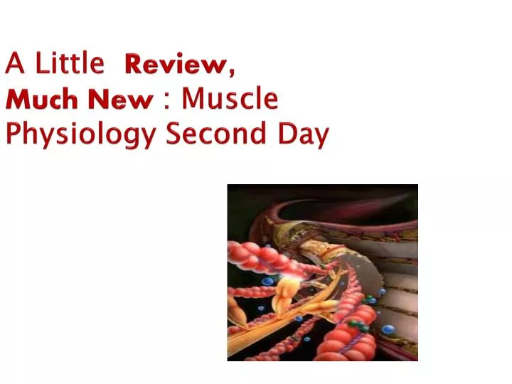
A Little Review , Much New : Muscle Physiology Second Day . Let’s Review Muscle Anatomy. Can you identify the parts of the muscle diagram on the left?. Characteristics of Muscle. Responsiveness (excitability) to chemical signals, stretch and electrical changes across the plasma membrane

E N D
Let’s Review Muscle Anatomy • Can you identify the parts of the muscle diagram on the left?
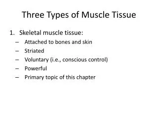
Characteristics of Muscle • Responsiveness (excitability) • to chemical signals, stretch and electrical changes across the plasma membrane • Conductivity • local electrical change triggers a wave of excitation that travels along the muscle fiber • Contractility -- shortens when stimulated • Extensibility -- capable of being stretched • Elasticity -- returns to its original resting length after being stretched
Remember what we say in A&P: FORM IS FOR FUNCTION!
IN Muscles, We See This Phenomenon especially well .. On The CELLULAR LEVEL
Generalized Animal Cell Figure 3.4
The Muscle Cell: A Specialized Cell: Called a myocyte, but better term is… Muscle Fiber.
Compared to a “typical” Cell The Muscle Fiber is : • Enormous!: Could have a diameter of 100um and can be length of a tendon ( up to 30 cm) • They are MULTINUCLEATE (a result of embryonic development…several myoblasts fuse together…each myoblast contributes a nucleus to the myofiber. • Satellite cells are unspecialized myoblasts that remain in myofiber. They are used for repair of damaged muscle fibers. • Glycogen; This carbohydrate is abundant in myofiber. • Myoglobin: Supplimental blood Pigment that carries oxygen to mitochondria under conditions of lower oxygen (hypoxia).
In the Muscle Fiber we Can See… • Myofibrils-long protein bundles( these are the muscle fibers) • The SR (Sarcoplasmic Reticulum); The function of this organelle is to act as reservoir of Calcium ions. SR- forms a network around each myofibril and exhibits dilated end-sacs called CISTERNAE. “Sister-NAY” • Transverse Tubules (T-Tubules): These will signal SR when to release Calcium into the cytosol. • Mitochondria- plenty of these ( in between the myofibrils) • Nuclei- Several, flattened against the sarcolemma.
The Muscle Cell: A Specialized Cell: Called a myocyte, but better term is… Muscle Fiber.
Myoblasts These are the embryonic stem cells that fuse during development to form the myofiber. They each contribute a nucleus. Some of them do not fuse, however. These “cells” that reside on the myofiber are called satellite cells. After an injury they assist in repair.
Quiz Yourself: • Why would muscle cells need an additional oxygen-carrying pigment ( myoglobin)? • Why is there a carbohydrate source readily available in the myofiber? • What is the difference between a myofiber and a myofibril?
Myofibrils Each myofibril is composed of 2 types of myofilaments: THIN filaments …..actin and THICK filaments…..myocin.
The A Band • M-line: Dark staining proteins found here in the middle of the H section. It keeps the myofibrils held together. • H –Zone: In a resting sarcomere, it contains ONLY thick filaments, and is the lighter region on sides of M line. • Zone of Overlap: In a resting sarcomere, thin filaments are situated between thick filaments.
The I band • Each I band contains thin filaments only and extends A band of one sarcomere to A band of next sarcomere. • HOWEVER, the Z Line marks the end of each sarcomere. • Remember, myofibrils consist of multiple sarcomeres that run adjacently. • Although most of the THICK MYOCIN is NOT in I band …Thin strands linger out …as a protein called TITAN to CONNECT to the Z line. ATTACHMENT SITE!!!
Striations = Organization of Filaments • Dark A bands (regions) alternating with lighter I bands (regions) • anisotrophic (A) and isotropic (I) stand for the way these regions affect polarized light • A band is thick filament region • lighter, central H band area contains no thin filaments • I band is thin filament region • bisected by Z disc protein called connectin, anchoring elastic and thin filaments • from one Z disc (Z line) to the next is a sarcomere
Thick Filaments • Made of 200 to 500 myosin molecules • 2 entwined polypeptides (golf clubs) • Arranged in a bundle with heads directed outward in a spiral array around the bundled tails • central area is a bare zone with no heads
Thin Filaments • Two intertwined strands fibrous • Groove holds tropomyosin molecules • One small, calcium-binding troponin molecule on each tropomyosin molecule
NOW, Arrange the Filaments • The thick and thin filaments of myofibrils are organized into REPEATING UNITs, often 10,000 of them end to end this is the ………….SARCOMERE
Muscle Contraction Begins When.. • Stored Calcium is released into the sarcoplasm • These ions are released into the sarcomeres
Relaxed and Contracted Sarcomeres • Muscle cells shorten because their individual sarcomeres shorten • pulling Z discs closer together • pulls on sarcolemma • Notice neither thick nor thin filaments change length during shortening • Their overlap changes as sarcomeres shorten
So The Sarcomere Has ALL the Structure It Need To Contract.. • NOW We Need to Begin To Discuss the Mechanism of Stimulating that Contraction….
What is ACTUALLY happening when a Sacromere Shrinks? • The thin filament ACTIN and the THICK filament MYOCIN form a CROSS-BRIDGE that cause a SLIDING motion. • This shortens the sacromere or closes that GAP or I band and causes a CONTRACTION. • Neither the ACTIN nor the Myocin filament actually shorten its the sacromere itself.
So …..HOW DO WE GET THE CROSS-BRIDGE BETWEEN ACTIN AND MYOSIN???
ACTIN and MYOSIN MUST BIND TO FORM a CROSS-BRIDGE BUT HOW?????? • First we know about the utrastructure of ACTIN filament and its PROTEINS.
ACTIN’s Ultrastructure • Fibrous F-Actin- This is the double-stranded protein (looks like a beaded necklace). • Globular G-Actin- These are the subunits of F-actin- contain active sites for myocin heads • Topomyosin- This is the filament that blocks the myosin sites. • Troponin- Found on tropomyosin, this protein binds w/ Ca++ . When it does, the configuration of tropomyosin will change…..the sites for myosin on ACTIN will become EXPOSED.
Classification of these Proteins? • Actin and Myosin ( CONTRACTILE PROTEINS..because they do the work of shortening the muscle fiber) • Tropomyosin and Troponin (Regulatory proteins- because they are like a switch that can be turned on and off.)
A Series of Events Must Occur Before Ca++ is Bound to Troponin: Let’s Begin • The control of the CONTRACTION is derived from the NERVOUS SYSTEM • The specialized intercellular connection between the nervous system and the muscle is known as the NEUROMUSCULAR JUNCTION
AND THAT IS THE TOPIC OF OUR NEXT LECTURE….. • UNTIL THEN….REVIEW THESE SLIDES, REREAD THE SECTIONS IN YOUR TEXT AND REVIEW YOUR LAB MANUAL PAGES ON MUSCLE PHYSIOLOGY..