1 / 37
2 likes | 21 Views
The bone of the skeleton is a mineralized vascular type of connective tissue with a solid matrix. The alveolar process is the bony extension of the mandible and maxilla that provides the necessary support for the teeth and serves as a site of attachment for the periodontal ligament fibers. By its resorption and deposition, it also compensates for tooth movement.

E N D
ALVEOLAR BONE ALVEOLAR BONE By Prof. Olfat M. Gaballah
CONTENTS CONTENTS • Classification of bone • Function of bone • Composition of bone • Cells of the bone • Alveolar process • Internal reconstruction of bone • Clinical considerations
Classification of Bone Classification of Bone Bone is a living mineralized connective tissue of the body, which serves various functions. • It forms the skeleton of the body. • It protects the vital organs (e.g. brain and lungs). • It acts as a reservoir for calcium. • It has a role as the load-bearing structure of the body. • Classification of bone Shape : Long (femur , tibia , fibula , ulna ,------) Short ( locate in the ankle and wrist joints ) Flat ( in skull ( parietal , occipital , frontal ( ( ribs , sternum ….) Irregular bone vertebrae , mandible, maxilla , zygoma -------- Development: Endochondral ( ossification occurs within the cartilaginous template mainly ) Intramembranous (ossification occurs direct in mesenchymal template ) Microscopic structure: compact bone, cancellous bone woven bone.
Chemical Composition of Bone Chemical Composition of Bone
Chemical composition of bone Chemical composition of bone • By weight, bone consisting of about : • 33% organic substance, and 67% inorganic material. • The organic substance, consists of 28% type I collagen and 5% noncollagenous proteins. • Noncollagenous proteins are bone sialoprotein, osteocalcin, osteonectin, osteopontin, and proteoglycans; growth factors and serum proteins also are found in bone. • The inorganic material is hydroxyapatite (Ca10 [PO4]6[OH]2).
Cells of the bone Cells of the bone Osteoprogenetors cells: undifferentiated mesenchymal cells that can differentiate into osteoblasts. Osteoblasts: these are mononucleated cells that synthesize and secrete the bone matrix Osteocytes: These are mature cells that lie within the fully formed bone and that no longer secret matrix. Each cell occupies a small chamber called a lacuna. The osteocytes derive from osteoblasts. Osteoclasts: these are cells responsible for the resorption of bone.
Osteoblasts: Osteoblasts are easily recognized in light microscopic sections as cuboid cells with open-faced nuclei and abundant basophilic cytoplasm. The cell membrane has a few cytoplasmic processes. -By electron microscopy, active osteoblasts contain a well developed rough endoplasmic reticulum, extensive Golgi apparatus and secretory vesicles and organelle that typically of protein secreting cells. Osteoblasts are mononucleated cells that synthesize both collagenous and noncollagenous bone matrix proteins such as bone sialoprotein and osteopontin. Some of these constituents first accumulate as an uncalcified matrix called osteoid that is composed mainly of collagen that will act as a scaffold for the deposition of the apatite crystals of bone. Osteoblasts are also responsible for the mineralization of bone as they exhibit high levels of alkaline phosphatase on their plasma membrane When bone is no longer forming, osteoblasts flatten substantially, extending along the bone. These cells termed bone lining cells,
Osteocytes: These cells are mature flat bone cells surrounded by bone matrix. They developed from imprisoned osteoblasts. They are incorporated in their surrounding matrix and have several branching processes. Each cell body is surrounded by a space of lacuna from which canaliculi arise that contains the processes of the cells and connect neighboring osteocytes together and with the osteoblasts or lining cells on the bone surfaces, the endosteum, periosteum and haversion canals, through gap junction forming osteoblasts-osteocytes complex it is necessary to bone matrix maintenance and vitality.
Osteoclasts Osteoclasts They are large cells having 15-20 nuclei. Derived from monocytes – macrophages Have a ruffled border on the cell surface. Always found in concave areas known as “Howshipslacunae” The ruffled border is adjacent to bone surface and it is surrounded by a clear zone that has only fine granular cytoplasm with microfilaments. The clear zone or sealing zone attaches the cells to the mineralized surface and sealing the periphery of the ruffled border) isolates a microenvironment between them and the bone surface.
Osteoclasts have a foamy eosinophilic cytoplasm and are rich with acid phosphatase and other lysosomal enzymes. The cell organelles consist of many nuclei, each of which is surrounded by multiple Golgi complexes, mitochondria, rough endoplasmic reticulum, and numerous vesicular structures situated between the Golgi complex and resorption surface. Another feature of osteoclasts is a proton pump associated with the ruffled border that pumps hydrogen ions into the sealed compartment
The sequence of resorptive events Attachment of osteoclasts to the mineralized surface of bone. Creation of a sealed acidic microenvironment through the action of the proton pump, which demineralizes bone and exposes the organic matrix. Secretion of organic acids mainly citric acid and lactic acids that chelate bone and increase the solubility of hydroxyapatite. Degradation of the exposed matrix by the action of released enzymes such as acid phosphatase and cathepsin B. Collagenase enzyme is secreted by osteoclasts and breaks down the collagen fibers extracellular. Endocytosis at the ruffled border of organic degradation products Translocation of degradation products in transport vesicles and extracellular release along the membrane opposite the ruffled border (transcytosis )
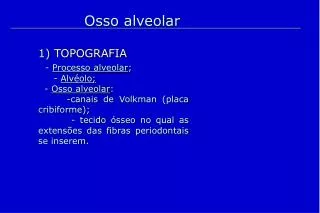
Alveolar bone (process) Alveolar bone (process) Definition: the alveolar process is that part of the maxilla and mandible that forms and supports the sockets of teeth and crypts of developing teeth
Development of alveolar bone Development of alveolar bone Development: it starts in the second month of fetal life The maxilla and mandible form a groove that is open towards the surface of the oral cavity. Developing tooth buds are enclosed in these grooves. Major portion of the alveolar process begins with root formation and eruption of the teeth. • During the rapid growth of alveolar bone, which occurs during fetal life areas of secondary cartilage may appear at the growing alveolar margins. • After the eruption of teeth, the alveolar bone gradually takes out its adult form. • As a result of its adaptation to function two parts of the alveolar bone can be distinguished: • 1- The alveolar bone proper. • 2- The supporting alveolar bone.
Structure of alveolar bone Structure of alveolar bone ALVEOLAR BONE ALVEOLAR BONE PROPER SUPPORTING ALVEOLAR BONE CORTICAL PLATE LAMELLATED BONE BUNDLE BONE SPONGY BONE
Woven Bone: • The Woven bone is immature bone or primitive bone. • It is characterized by a great number, great size and irregular arrangement of osteocytes. • Its collagen fibers are irregularly arranged. The great amount of organic substance and the reduced volume of inorganic material render this bone more radiolucent than mature bone. • This bone is finally resorbed and replaced by lamellar bone
The Basal Bone • The basal bone can be defined as that, it is the bone of the facial skeleton that supports the alveolar bone. Both the alveolar process and basal bone are covered by the same periosteum.
Periosteum and endosteum Periosteum and endosteum • Periosteum: identifiable on the outer surface of the bone. It consists of two layers; the Outer fibrous membrane layer and the inner cellular layer. • The Outer firm fibrous layer made up of collagen and reticular fibers, blood vessels, and nerves • The inner layer is an osteogenic layer. presents adjacent to bone and contains smaller blood vessels and proliferative cells of bone. • Endosteum: A membrane lining the Bone marrow cavity, the Haversian canal, and all the internal cavities of the bone. The endosteum consists of a layer of osteoprogenitor cells and collagenous fibers The endosteum is noticeably thinner than the periosteum.
Alveolar Bone Proper • Alveolar bone proper consists of a thin lamella of bone that surrounds the roots of the teeth and gives attachment to the principal fibers of the periodontal ligament. • It is perforated by many openings that carry branches of the intra alveolar nerves and blood vessels into the periodontal ligament, and is called the cribriform plate. • It consists of bundle bone Lamellated bone Lamellated bone consists of Concentric lamellae along with a central blood vessel form an osteon. • NOTE : Interdental and interradicular septa have canals known as canals of “ zukerkandl and hirschfeld”
Bundle bone: Bundle bone: It is that part of the alveolar process into which the fiber bundles of the periodontal ligament insert Histologically : the term bundle bone is chosen because --- the bundles of the principal fibers of the periodontal ligament continue into the bone as Sharpey`s fibers Sharpey’s fibres are seen perpendicular to the bundle bone. Radiographic: it is named lamina dura. It appears more radiopaque due to the presence of thick bone without trabeculations Clinically: The lamina dura is an important diagnostic landmark in determining the health of the periapical tissues. Loss of density usually means a pathological condition
Supporting Alveolar Bone • It consists of two parts – • Cortical plates (Outer and inner) • Spongy bone Cortical plates: These are made up of compact bone & form the outer and inner plates of the alveolar bone. Cortical bone varies in thickness in different areas. it is thicker in the mandible than in the maxilla and thicker in the premolar-molar region than in the anterior. it consists of Haversion system which is formed of Haversion canals that run parallel to the long axis of the bone and contain blood vessels. -Osteocytes in their lacunae are present -Volkmann's canals connect the haversioncanals with the external surface of the bone and the marrow spaces. While the transverse canals connect between two haversioncanals
• Spongy Bone: • it fills the area between the cortical plates and the alveolar bone proper. • It contains trabaculaeof bone and marrow spaces. • (It is formed of trabeculae of the bone surrounding the medullary spaces that contain the bone marrow. The trabeculae are made up of varying numbers of closely adjoining lamellae with lacunae and osteocytes in between). • Types of spongy bone (spongiosa) :- • Type I: the trabeculae are regular and horizontal like a ladder. It is seen most commonly in the mandible. • Type II: irregularly arranged delicate and numerous trabeculae. It is seen most commonly in the maxilla. The spongy bone is very thin or absent in the anterior regions of both jaws. • Around functionless teeth, the spongy bone shows very wide medullary spaces and little number of trabeculae.
Spongy bone Spongy bone
SPONGY bonev COMPACT bone
Alveolar Crest • The alveolar crest: is the most coronal portion, or the top, of the alveolar process. The alveolar crest is often the first portion of the alveolar process that is damaged by periodontal disease and is therefore the first bone that is lost. • The shape and position of the crest are dependent on the adjacent teeth. The alveolar crest is around 1.5 to 2 mm below the cementoenamel junction in a healthy mouth. •
Internal reconstruction of bone Internal reconstruction of bone Bone is a dynamic tissue and is always undergoing changes to adapt to functional forces, mesial drift, and eruption of teeth. There is constant formation and resorption of bone. Periods of resorption alternate with periods of rest and repair. Incremental lines of bone Faint line • These are formed by the rhythmic apposition of bone during bone development. • This rhythmic apposition, is due to a change of direction of the collagen fibers ( 45 degree ) of matrix from one layer to another. Resting line: these lines correspond to the resting period in the process of continuous bone formation. Reversal line: when a period of bone resorption is followed by bone formation, a dark line is seen which separates the new bone from the old bone, . It is scalloped in shape with the convexity toward the old bone
Incremental lines of Bone Incremental lines of Bone
Clinical Considerations Bones are not inert structures within the human body; they continue to change over the course of a lifespan. Through bone remodeling the alveolar bone proper may become displaced in relation to the remaining alveolar process, thereby allowing tooth movement to take place. Interruptions in the continuity of the lamina dura in the apical region of an alveolus are of diagnostic significance in the radiographic identification of periapical lesions. Proximity of the alveolar bone to sinus cavities or major nerves (mandibular nerve) may create problems during tooth extraction or surgical interventions. Following tooth extraction, the alveolar process tends to resorb.(Lack of mechanical stimulation: After a tooth is extracted, the normal mechanical stimulation from chewing and biting that the tooth provided to the surrounding bone is lost. This lack of loading on the bone can lead to resorption. Placement of dental implants in the alveolar process, prior to its becoming resorbed, following tooth extractions will markedly decrease the rate of ridge resorption
References Nanci a: wheeler's dental anatomy, physiology, and Occlusion. 11th edition - November 9, 2019 Kumar GS: Orban’s oral histology and embryology. 15Th Edition 2019 Avery: essentials of oral histology and embryology. A Clinical approach, 3rd edition, elsevier's publisher, Philadelphia. http://www.annclinlabsci.org/content/5/4/264.full.pdf https://2u.pw/xp4NAbu https://2u.pw/kJy9Nvv