Download

1 / 24
240 likes | 1.4k Views
How to detect coronary heart disease. If the patient does not have chest pain ... Carotid IMT and future heart attacks and strokes in individuals with no symptoms ...

E N D
Causes of death in the USA • Heart disease is number one • Newsweek, Jan 31 2000 (p. 49)
Where does heart disease come from ? • Epidemiologic studies, e.g. the Framingham Heart Study • Linkage between “risk factors” and coronary artery stenosis • What is a risk factor ? • Elevated blood pressure • Elevated cholesterol • Cigarette smoke
How to detect coronary heart disease • If the patient has chest pain • Stress testing • Radionuclide imaging • Coronary catheterization • This is for “advanced disease”
How to detect coronary heart disease • If the patient does not have chest pain • This is for early, possibly reversible disease
MESAMulti-Ethnic Study of Atherosclerosis • NHLBI/NIH funded • Multi-center study:6 centers, 6500 participants • Risk factors for atherosclerosis • Non-invasive markers of atherosclerosis • Coronary calcium scoring • Progression of calcium scores
MESAMulti-Ethnic Study of Atherosclerosis • Ultrasound markers of atherosclerosis • Carotid IMT • Brachial artery reactivity (endothelial function) • Carotid artery distensibility
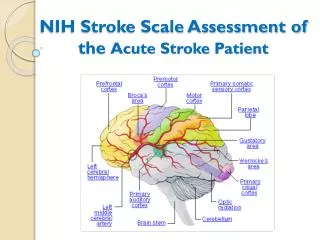
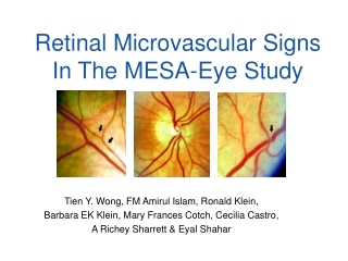
What is Carotid IMT ? IMT: thickness of the intima to media interfaces seen on high resolution imaging of the arterial wall
40 30 20 10 0 1 2 3 4 5 Carotid IMT and future heart attacks and strokes in individuals with no symptoms Top 20% with thick IMTs Combined IMT CCA IMT 29 29 28 ICA IMT 5 times the chance of heart attack and stroke 21.2 20.6 19.5 18.1 Incidence Rates (1,000 person-years) 16.1 16 12.8 12.2 10.4 6.2 5.6 4.8 O’Leary, Polak, Kronmal et al. NEJM 1999 Quintiles of IMT values
Coronary artery physiology • Healthy coronary arteries should dilate when needed • Coronary reactivity is abnormal in patients with coronary disease • May represent a generalized dysfunction of the arterial system
Brachial artery reactivity • Brachial artery endothelial function parallels coronary response • Abnormal: less of an increase in diameter during stimulus
MESA: Ultrasound device selection • Goal: select an imaging device that will permit precise measurements of: • Level of wall thickening in the carotid artery: IMT • Changes in arterial diameter over time: brachial artery reactivity
MESA: Ultrasound device selection • Acuson Aspen, Hewlett-Packard ImagePoint; Siemens Allegra; Toshiba Power Vision 8000; GE Logiq-700; ATL HDI-5000 • Same sonographer, same subject • Operator: application specialist from the respective companies
MESA: Ultrasound device selection • 2 of the 6 devices disqualified • Poor image quality • Insufficient frame rates • Unanimous decision by 6 representatives of the field centers
MESA: Ultrasound device selection • Removal of identifying marks • Scaling to same magnification • Normalized image intensities (0 to 255; minimum to maximum) • Scoring as 1 (best) to 4 (worse) • Images transmitted by e-mail
MESA: Ultrasound device selection • Image evaluation performed by: • 6 trained readers: perform the measurements • 6 representatives from the field centers • Scores tallied for • Carotid IMT near wall • Carotid IMT far wall • Brachial artery image clarity
MESA: Ultrasound device selection • Brachial artery diameters
MESA: Ultrasound device selection • Carotid IMT far wall
MESA: Ultrasound device selection • Device ranking
MESA: Ultrasound device selection • Device ranking
MESA: Ultrasound device selection • Based on device ranking, the GE Logiq-700 was picked for MESA • The study has enrolled more than 2000 of 6500 individuals • Preliminary examination of data quality and completeness shows overall good performance for all imaging tasks
MESA: Ultrasound examination • Unanswered questions: • Are coronary artery calcium scores superior to carotid IMT ? • Does endothelial dysfunction predict the development of coronary disease ?