Download
1 / 12
120 likes | 523 Views
Lecture 15. Sense Organs I: The Visual System. Visual System. Eye Accessory structures Eyebrows, eyelids, eyelashes, lacrimal (tear) glands Optic nerve (II) Sensory information travels along optic nerve to thalamus then to occipital lobe. Accessory Structures of Eye. Eyebrows

E N D
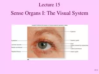
Lecture 15 Sense Organs I: The Visual System
Visual System • Eye • Accessory structures • Eyebrows, eyelids, eyelashes, lacrimal (tear) glands • Optic nerve (II) • Sensory information travels along optic nerve to thalamus then to occipital lobe
Accessory Structures of Eye • Eyebrows • Prevent running perspiration into eyes • Shade • Eyelids or palpebrae • Protect and lubricate • Lacrimal caruncle has sebaceous and sweat glands • Extrinsic eye muscles (6) • Lacrimal apparatus • Lacrimal gland (facial nerve VII) • Conjunctiva • Thin mucous membrane over inner surface of eyelids and anterior side of eyeball Fig. 19.10
Anatomy of the Eye • Three coats or tunics • Fibrous: Consists of sclera and cornea • Vascular: Consists of choroid, ciliary body, iris • Neural: Consists of retina Fig. 19.12
Fibrous tunic: Outer Sclera: White outer layer, maintains shape, protects internal structures, provides muscle attachment point, continuous with cornea Cornea: No blood vessels, transparent, allows light to enter eye and refracts (bends) light Anatomy of the Eye Fig. 19.12
Vascular tunic: Middle Iris: Controls light entering pupil; smooth muscle Ciliary muscles: Control lens shape; smooth muscle Choroid: pigmented layer that contains melanin Anatomy of the Eye
Retina: Inner Contains neurons sensitive to light Macula lutea & Fovea centralis: Area of greatest visual acuity Optic disc: Blind spot Cavities Anterior: Aqueous humor Posterior: Vitreous humor Anatomy of the Eye
Lens Held by suspensory ligaments attached to ciliary muscles Transparent, biconvex Anatomy of the Eye
Focus and Accommodation • Far vision: 20 feet or more from eye • Near vision: Closer than 20 feet • Accommodation Fig. 19.16
Review Question Damage to the retina due to excessive light entering the eyeball could indicate a problem with the • Iris • Ciliary body • Cornea • Conjunctiva • Choroid
Points to Remember • Accessory eye structures protect and lubricate the eye. • There are 3 basic layers to the eye: fibrous tunic, vascular tunic and neural tunic. • Image formation on retina by bending of light rays through cornea and lens. • To view close objects, the lens increases its curvature.