Download
1 / 79
820 likes | 2.08k Views
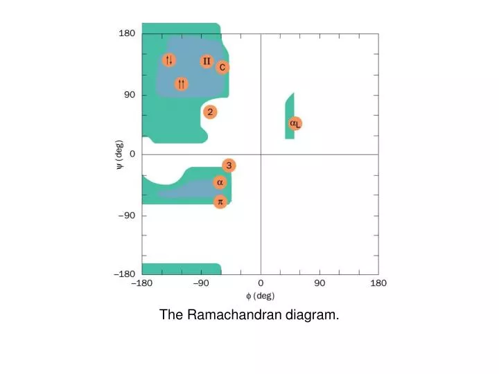
The Ramachandran diagram. Allowed phi and psi torsion angles in proteins. The Ramachandran diagram of Gly residues in a polypeptide chain. Cis/Trans Isomerization: Proline. trans. cis. Energy difference between these forms is small.

E N D
The Ramachandran diagram of Gly residues in a polypeptide chain.
Cis/Trans Isomerization: Proline trans cis Energy difference between these forms is small. Nearly all Xaa-Pro linkages are biosynthesized in the trans form. ~10% of these peptide bonds are in the cis form in globular proteins. Interconversion catalyzed by peptidyl prolyl cis-trans isomerases
Stereo space-filling representation of an a helical segment of sperm whale myoglobin (its E-helix) as determined by X-ray crystal structure analysis.
The hydrogen bonding pattern of several polypeptide helices.
Comparison of the two polypeptide helices that occasionally occur in proteins with the commonly occurring a helix.
A two-stranded b antiparallel pleated sheet drawn to emphasize its pleated appearance.
Stereo space-filling representation of the 6-stranded antiparallel b pleated sheet in jack bean concanavalin A as determined by crystal X-ray analysis.
Polypeptide chain folding in proteins illustrating the right-handed twist of b sheets: bovine carboxypeptidase A.
Polypeptide chain folding in proteins illustrating the right-handed twist of b sheets: chicken muscle triose phosphate isomerase. (b barrel)
hairpin out-of-plane crossovers Connections between adjacent polypeptide strands in b pleated sheets.
Space-filling representation of an Ω loop comprising residues 40 to 54 of cytochrome c.
The two-stranded coiled coil: view down the coil axis showing the interactions between the nonpolar edges of the a helices.
The two-stranded coiled coil: side view in which the polypeptide back bone is represented by skeletal (left) and space-filling (right) forms.
The amino acid sequence at the C-terminal end of the triple helical region of the bovine a1(I) collagen chain.
X-Ray structure of the triple helical collagen model peptide (Pro-Hyp-Gly)10 in which the fifth Gly is replaced by Ala. (a) Ball and stick representation.
X-Ray structure of the triple helical collagen model peptide (Pro-Hyp-Gly)10 in which the fifth Gly is replaced by Ala. (b) View along helix axis.
X-Ray structure of the triple helical collagen model peptide (Pro-Hyp-Gly)10 in which the fifth Gly is replaced by Ala. (c) A schematic diagram.
A biosynthetic pathway for cross-linking Lys, Hyl, and His side chains in collagen.
X-Ray diffraction photograph of a single crystal of sperm whale myoglobin.
Electron density maps of proteins (heme of sperm whale myoglobin) (2 angstrom resolution)
Electron density maps of proteins (sperm whale myoglobin)(2.4 angstrom resolution)
Sections through the electron density map of diketopiperazine calculated at the indicated resolution.
The 2D proton NMR structures of proteins: a NOESY spectrum of a protein presented as a contour plot with two frequency axes w1 and w2.
The 2D proton NMR structures of proteins: NMR structure of a 64-residue polypeptide comprising the Src protein SH3 domain.
Representations of the X-ray structure of sperm whale myoglobin: the protein and its bound heme are drawn in stick form.
8 helices Representations of the X-ray structure of sperm whale myoglobin: a diagram in which the protein is represented by its computer-generated Ca backbone.
Representations of the X-ray structure of sperm whale myoglobin: a computer-generated cartoon drawing.
The x-ray structure of horse heart cytochrome c. (hydrophobic residues in red)
The x-ray structure of horse heart cytochrome c. (hydrophilic residues in green)
H-helix Representations of the x-ray structure of sperm whale myoglobin: a diagram in which the protein is represented by its computer-generated Ca backbone.
The H helix of sperm whale myoglobin. (a)A helical wheel representation in which the side chain positions about the a helix are projected down the helix axis onto a plane.
The H helix of sperm whale myoglobin: a skeletal model.(orange = nonpolar; purple = polar)
orange = nonpolar purple = polar The H helix of sperm whale myoglobin: a space-filling model.
red = nonpolar purple = polar A space-filling model of an antiparallel b sheet from concanavalin A.
two domains One subunit of the enzyme glyceraldehyde-3-phosphate dehydrogenase from Bacillus stearothermophilus.
aa bab b-hairpin Schematic diagrams of supersecondary structures.
Greek key motif Schematic diagrams of supersecondary structures.
directionality of helices X-ray structures of 4-helix bundle proteins: E. coli cytochrome b562.
directionality of helices X-ray structures of 4-helix bundle proteins: human growth hormone.
stacked 4-stranded and 3-stranded antiparallel b-sheets X-ray structure of the immunoglobulin fold.