Download
1 / 28
350 likes | 835 Views
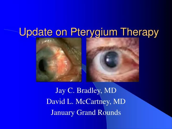
Update on Pterygium Therapy. Jay C. Bradley, MD David L. McCartney, MD January Grand Rounds. From the BCSC: Basics. Often bilateral Almost always situated at the nasal or temporal limbus within palpebral fissure Associated with prolonged UV exposure

E N D
Update on Pterygium Therapy Jay C. Bradley, MD David L. McCartney, MD January Grand Rounds
From the BCSC: Basics • Often bilateral • Almost always situated at the nasal or temporal limbus within palpebral fissure • Associated with prolonged UV exposure UV-B limbal stem cell p53 mutation apoptosis / TGF- growth • May be associated with dryness, inflammation, and exposure to wind and dust or other irritants • Prevalence increases with proximity to equator • Difficult to determine if race is independent risk factor due to confounding variables
Albedo Hypothesis • Researcher: MT Coroneo (Australia) • Pterygia occur secondary to albedo concentration in the anterior eye • Light entering the temporal limbus at 90 degrees is concentrated onto the medial limbus • Related to corneal curvature • Explains predominance of medial pterygia Ophthalmic surg. 1990 Jan;21(1):60-6.
From BCSC: Basics • Encroaches on cornea in wing-like fashion • Overlying epithelium often thinned, but can be hyperplastic or dysplastic • Nearly always preceded by pingueculae • Induces astigmatism (usually “with-the-rule”) proportional to size • Excision indicated if persistent irritation, vision distortion, significant (> 3-4 mm) and progressive growth toward visual axis, restricted ocular motility, and atypical appearance
From the BCSC: Basics • Elastotic degeneration – fragmentation and breakdown of stromal collagen • Destruction of Bowman’s layer by advancing fibrovascular tissue resulting in corneal scarring
From BCSC: Basics • Recurrent pterygia – lack elastotic degeneration and are more accurately classified as an exuberant granulation tissue response • Stocker’s line – a pigmented iron line in advance of pterygium
Pterygium Excision • Goal: Achieve a normal, topographically smooth ocular surface • Dissect a smooth plane toward the limbus • Some surgeons prefer specialized blunt pterygium blades (Tooke or Gills) while others prefer sharp blades • Preferable to dissect down to bare sclera at limbus • Bare sclera = remove loose Tenon’s layer and leave episcleral vessels intact
Some surgeons avoid medial dissection to avoid bleeding from trauma to adjacent muscle tissue while other remove excessive fibrovascular tissue medially • Light thermal cautery is applied for hemostasis
Pterygium Recurrence • Growth of fibrovascular tissue across the limbus onto cornea after initial removal • Excludes persistence of deeper corneal vessels and scarring which may remain even after adequate removal • Bunching of conjunctiva and formation of parallel loops of vessels, which aim almost like an arrowhead at the limbus, usually denotes a conjunctival recurrence
Proposed Recurrence Grading System • Grade 1 – normal appearing operative site • Grade 2 – fine episcleral vessels in the site extending to the limbus • Grade 3 – additional fibrous tissues in site • Grade 4 – actual corneal recurrence
Wound Closure Options: • Bare sclera • Simple closure • Sliding flap • Rotational flap • Conjunctival graft
Bare Sclera Closure • No sutures or fine, absorbable sutures used to appose conjunctiva to superficial sclera in front of rectus tendon insertion • Leaves area of “bare sclera” • Relatively high recurrence rate with variable techniques of 5 – 68 % with primary / 35 – 82 % with recurrent)
Simple Closure • Free edges of conjunctiva secured together • Effective only if defect is very small • Can be used for pingueculae removal • Reported recurrence rates from 45 – 69 % (one report of “barest” sclera, N=800 of 2 %) • Few complications (dellen)
Sliding Flap Closure • An L-shaped incision is made adjacent to the wound to allow conjunctival flap to slide into place • Reported recurrence rates from 0.75 – 5.6 % (poorly designed, retrospective) • Few complications (flap retraction / cyst formation)
Rotational Flap Closure • A U-shaped incision is made adjacent to the wound to form tongue of conjunctiva that is rotated into place • Reported recurrence of 4 % • Few complications
Conjunctival Graft Closure • A free graft, usually from superior bulbar conjunctiva, is excised to correspond to wound and is then moved and sutured into place • Can be performed with inferior conjunctiva to preserve superior conjunctiva
Conjunctival Graft Closure • Harvested tissue should be approximately 0.5 – 1 mm larger than defect • Most important aspect in harvesting is to procure conjunctival tissue with only minimal or no Tenon’s included • Graft is transferred to recipient bed and secured with or without incorporating episclera • Some surgeons harvest limbal stem cells along with graft and orient graft to place stem cells adjacent to site of corneal lesion excision
Conjunctival Graft Closure • Topical antibiotic-corticosteroid ointment used for 4 – 6 weeks post-operatively until inflammation subsides (compliance with this regimen decreases recurrence) • Used when extensive damage or destruction of limbal epithelial stem cells is NOT present • Reduces recurrence to 2 – 5 % (up to 40 % in some reports) • Ameliorates the restriction of extraocular muscle function
Limbal Conjunctival Autograft • Reported recurrence rates are variable (between 0 – 40 %) • Few complications • Further prospective studies in primary and recurrent pterygia are needed
Lamellar Corneal Transplant • Wound closed with piece of lamellar sclera or cornea • Reported recurrence rates of 6 – 30 % • Not performed often • Can be used in conjunction with AMT for multiply recurrent pterygia with corneal scarring and limited available conjunctiva • Method involves increased surgical complexity, the requirement of donor tissue, and risk of infectious disease transmission
Adjunctive Beta Irradiation • Most common dosage is 15 Gy in single or divided doses • Reasonably acceptable recurrence rates (from 0 – 50 % with bare sclera or simple conj closure) • Risk of corneal or scleral necrosis and endophthalmitis
Adjunctive Thiotepa • Most common dose is 1:2000 thiotepa given up to every 3 hours for approx. 6 weeks • Usually used with bare sclera method • Low reported recurrence rates of 0 – 16 % (poor study quality) • Minimal complications (2 cases of scleral thinning)
Adjunctive Mitomycin C • Used with bare sclera or conj closure • Most common dose is 0.02 % applied for 3 min during surgery • Risk of aseptic scleral necrosis / perforation and infectious sclerokeratitis • Used more often for recurrent cases • Rate of recurrence between 3 – 25 % for intra-op / 5 – 54 % for post-op with most studies showing < 10 % recurrence
Amniotic Membrane Graft Closure • Useful for very large conjunctival defects as in primary double-headed pterygium or to preserve superior conjunctiva for future glaucoma surgeries • Requires costly donor tissue • Reported recurrence rate between 3 – 64 % for primary cases and 0 – 37.5 % for recurrent cases
Other Methods: • Pterygium head transplantation • Split skin grafts • Ruthenium adjunctive therapy • Laser or thermal cautery • Excimer laser treatment • PDT (one report, N = 10) • Intraoperative doxorubicin / daunorubicin • 5-FU • Serum-free derived cultivated conjunctival graft • Recombinant epidermal growth factor ****Few studies with limited numbers of patients, poor follow-up, and variable recurrence rates
Primary Pterygium Metanalysis Includes 5 studies with N=290 (BS+Mito=257/CAG=33) ComparisonOdds Ratio95 % CI Bare sclera: mito C 25:1 9.0 – 66.7 Bare sclera: CAG 6:1 1.8 – 18.8 Sanchez-Thorin JC et al. Br J Ophthalmol 82:661-5, 1998.
Conclusions: • There is no clear-cut superior single treatment • Bare scleral and simple conjunctival closure without adjunctive therapy have relatively high but variable recurrence rates • Use of beta irradiation and antimetabolites can be used with appropriate caution • Conjunctival transplants and flaps appear to have overall lower rate of recurrence but require more surgical time and unnecessary conj destruction • Other treatment options need further adequate study prior to widespread implementation