Download
1 / 39

E N D
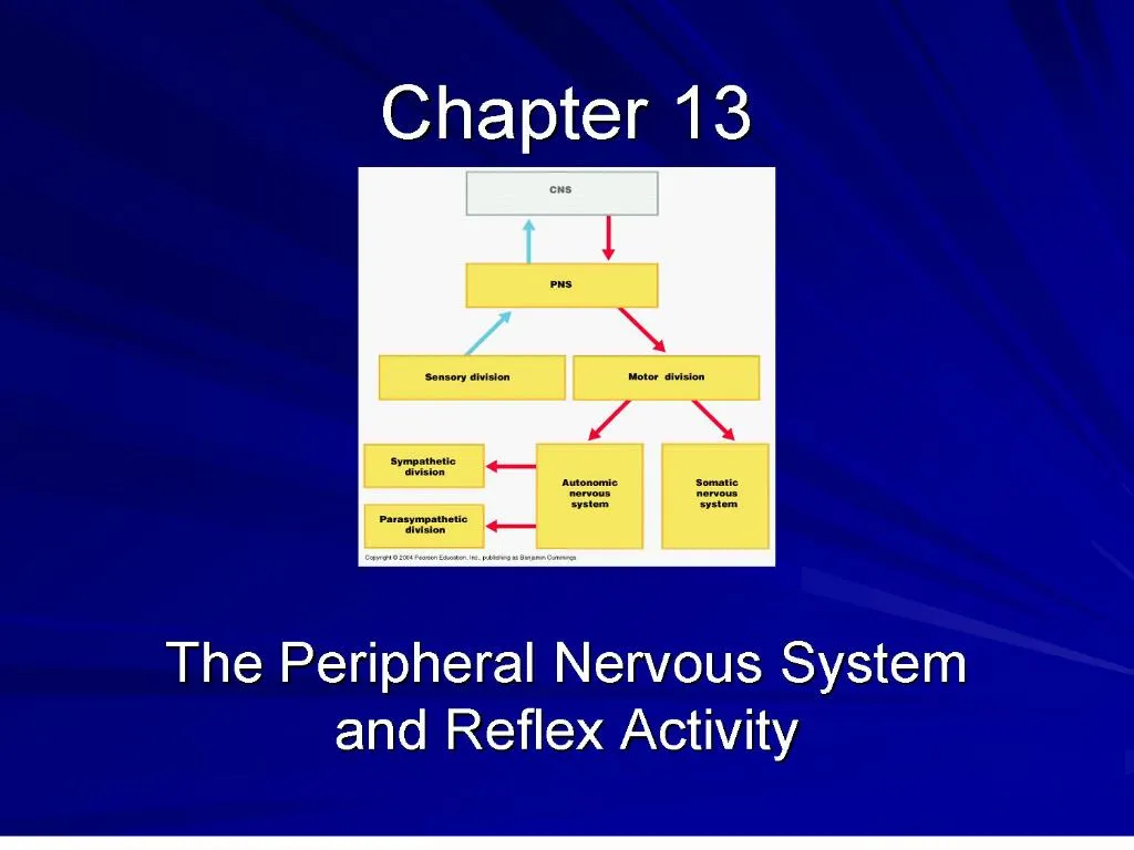
1. Chapter 13 The Peripheral Nervous System and Reflex Activity
2. Peripheral Nervous System PNS � all neural structures outside the brain and spinal cord
All neural structures outside the brain and spinal cord
Sensory receptors
Peripheral nerves and associated ganglia
Efferent motor endings
PNS � all neural structures outside the brain and spinal cord
3. Sensory Receptors Structures specialized to respond to stimuli
Activation of sensory receptors results in depolarizations that trigger impulses to the CNS
The realization of these stimuli, sensation and perception, occur in the brain
4. Receptor Classification by Stimulus Type Mechanoreceptors � respond to touch, pressure, vibration, stretch, and itch
Thermoreceptors � sensitive to changes in temperature
Photoreceptors � respond to light energy (e.g., retina)
Chemoreceptors � respond to chemicals (e.g., smell, taste, changes in blood chemistry)
Nociceptors � sensitive to pain-causing stimuli
5. Receptor Class by Location: Exteroceptors Respond to stimuli arising outside the body
Found near the body surface
Sensitive to touch, pressure, pain, and temperature
Include the special sense organs
6. Receptor Class by Location: Interoceptors Respond to stimuli arising within the body
Found in internal viscera and blood vessels
Sensitive to chemical changes, stretch, and temperature changes
7. Receptor Class by Location: Proprioceptors Found in skeletal muscles, tendons, joints, ligaments, and connective tissue coverings of bones and muscles
Respond to degree of stretch of the organs they occupy
Constantly �advise� the brain of one�s movements
8. Receptor Classification by Structural Complexity Receptors are structurally classified as either simple or complex
Most receptors are simple and include encapsulated and unencapsulated varieties
Complex receptors are special sense organs
9. Simple Receptors of the General Senses Involved in
Tactile sensation
Temperature monitoring
Pain
�Muscle sense�
Are either unencapsulated (free nerve endings) or encapsulated nerve endings
10. Unencapsulated Nerve Endings Free Nerve Endings
Located everywhere
Respond to pain, temperature, pressure
11. Unencapsulated Nerve Endings Merkel discs
Lie in deep layers of epidermis
Function as light touch receptors
12. Unencapsulated Nerve Endings Hair follicle receptors
Wrap around hair follicles
Respond to light touch
13. Encapsulated Nerve Endings Meissner�s corpuscles
Found just beneath the epidermis
Numerous on sensitive hairless areas
14. Encapsulated Nerve Endings Pacinian corupscles
Found everywhere
Stimulated by deep pressure
15. Encapsulated Nerve Endings Ruffini�s corpuscles
Lie deep in dermis, subcutaneous tissue and joint capsules
Respond to deep pressure and stretching
16. Encapsulated Nerve Endings Muscle spindles
Found in perimysia of skeletal muscles
Detect when a muscle is stretched
Initiated a reflex that resists the stretch
17. Encapsulated Nerve Endings Golgi tendon organs
Located in tendons, close to muscle insertion.
Stimulated when muscle stretches the tendon
18. Encapsulated Nerve Endings Joint kinesthetic receptors
Monitor stretch in articular capsules of synovial joints
Provide information on joint position and motion
20. Structure of a Nerve Nerve � cordlike organ of the PNS consisting of peripheral axons enclosed by connective tissue
Connective tissue coverings include:
Endoneurium � loose connective tissue that surrounds axons
Perineurium � coarse connective tissue that bundles fibers into fascicles
Epineurium � tough fibrous sheath around a nerve
22. Classification of Nerves Sensory and motor divisions
Sensory (afferent) � carry impulse to the CNS
Motor (efferent) � carry impulses from CNS
Mixed � sensory and motor fibers carry impulses to and from CNS; most common type of nerve
23. Regeneration of Nerve Fibers Damage is serious because neurons are amitotic
If the soma of a damaged nerve remains intact, damage can be repaired
Regeneration involves coordinated activity among:
Macrophages � remove debris
Schwann cells � form regeneration tube and secrete growth factors
Axons � regenerate damaged part
24. Cranial Nerves Twelve pairs of cranial nerves arise from the brain
They have sensory, motor, or both sensory and motor functions
Each nerve is identified by a number (I through XII) and a name
Four cranial nerves carry parasympathetic fibers that serve muscles and glands
25. Cranial Nerve I: Olfactory Origin: Receptors are in nasal cavity
Passes through the cribriform plate of the ethmoid bone
Fibers run through the olfactory bulb and terminate in the primary olfactory cortex
Functions solely by carrying afferent impulses for the sense of smell
26. Cranial Nerve II: Optic Origin: retina of the eye
Optic nerves converge at the optic chiasm
They continue to the thalamus where they synapse
From there, the optic radiation fibers run to the visual cortex
Functions solely by carrying afferent impulses for vision
27. Cranial Nerve III: Oculomotor Fibers extend from midbrain to extrinsic eye muscles
Functions in raising the eyelid, directing the eyeball, constricting the iris, pupil, and controlling lens shape
28. Cranial Nerve IV: Trochlear Fibers emerge from the dorsal midbrain and innervate the superior oblique muscle
Primarily a motor nerve that directs the eyeball
29. Cranial Nerve V: Trigeminal Fibers extend from pons to face and form
Mixed cranial nerve
3 divisions:
Ophthalmic division: sensory fibers from area surrounding eye
Maxillary division: sensory fibers from nasal cavity, upper teeth
30. Cranial Nerve V: Trigeminal Mandibular division: sensory fibers from tongue lower teeth, chin, and motor fibers to muscles of mastication
31. Cranial Nerve VI: Abducens Motor fibers run from pons to extrinsic muscle that abducts eye.
32. Cranial Nerve VII: Facial Fibers leave the pons, travel to the lateral aspect of the face
Mixed nerve with five major branches
Motor functions include facial expression, and the transmittal of autonomic impulses to lacrimal and salivary glands
Sensory function is taste from the anterior two-thirds of the tongue
33. Cranial Nerve VIII: Vestibulocochlear Fibers arise from the hearing and equilibrium apparatus of the inner ear and enter the brainstem at the pons-medulla border
Two divisions � cochlear (hearing) and vestibular (balance)
Functions are solely sensory � equilibrium and hearing
34. Cranial Nerve IX: Glossopharyngeal Fibers emerge from the medulla, leave the skull via the jugular foramen, and run to the throat
Mixed nerve with motor and sensory functions
Motor � innervates part of the tongue and pharynx, and provides motor fibers to the parotid salivary gland
Sensory � fibers conduct taste and general sensory impulses from the tongue and pharynx
35. Cranial Nerve X: Vagus The only cranial nerve that extends beyond the head and neck
Fibers emerge from the medulla via the jugular foramen
The vagus is a mixed nerve
Most motor fibers are parasympathetic fibers to the heart, lungs, and visceral organs
Its sensory function is in taste
36. Cranial Nerve XI: Accessory The accessory nerves are unique in that they are formed by the union of a cranial and spinal root
Primarily a motor nerve
Supplies fibers to the larynx, pharynx, and soft palate
Innervates the trapezius and sternocleidomastoid, which move the head and neck
37. Cranial Nerve XII: Hypoglossal Motor fibers arise from the medulla and innervates both extrinsic and intrinsic muscles of the tongue, which contribute to swallowing and speech
38. I. Olfactory
II. Optic
III. Oculomotor
IV. Trochlear
V. Trigeminal
VI. Aducens
On Occasion, Our Trusty Truck Acts Funny � Very Good Vehicle AnyHow
VII. Facial
VIII. Vestibulocochlear
IX. Glossopharyngeal
X. Vagus
XI. Accessory
XII. Hypoglossal
39. Some Say Money Makes Business, My Brother Says Big Business Makes Money


