Download
1 / 14
140 likes | 262 Views
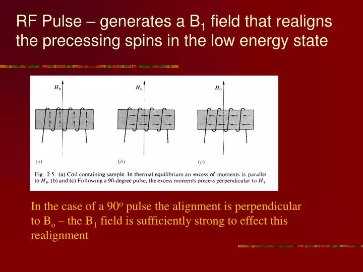
RF Pulse – generates a B 1 field that realigns the precessing spins in the low energy state . In the case of a 90 o pulse the alignment is perpendicular to B o – the B 1 field is sufficiently strong to effect this realignment. The NMR/MRI Signal and FT. Signal acquired by

E N D
RF Pulse – generates a B1 field that realigns the precessing spins in the low energy state In the case of a 90o pulse the alignment is perpendicular to Bo – the B1 field is sufficiently strong to effect this realignment
The NMR/MRI Signal and FT Signal acquired by the NMR/MRI unit Fourier Transform (FT) Line width 1/T2
Pulses, T1 and T2 • Pulses have nothing to do with either T1 or T2 • Relaxation mechanisms/processes pertain to the a) recovery of magnetization along the Bo or z-axis and/or b) the dephasing of the NMR/MRI signal in the x-y plane
The Importance of Relaxation Phenomena for fMRI and Clinical MRI • Small differences in T1 and T2 can provide clear-cut diagnostic interpretations • Small changes in signal intensity from the decrease of deoxyhemoglobin and the increase of oxyhemoglobin due to capillary dilation with brain activation are the basis for BOLD fMRI • MRI ‘contrast agents’ all rely on changing T1 or T2 of nearby water protons to give better disease changes contrast