Download
1 / 112
1.23k likes | 4.85k Views
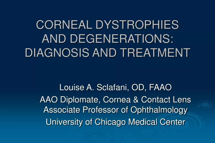
CORNEAL DYSTROPHIES AND DEGENERATIONS: DIAGNOSIS AND TREATMENT. Louise A. Sclafani, OD, FAAO AAO Diplomate, Cornea & Contact Lens Associate Professor of Ophthalmology University of Chicago Medical Center. GOALS. Differentiate dystrophy vs. degeneration Normal vs. abnormal

E N D
CORNEAL DYSTROPHIES AND DEGENERATIONS: DIAGNOSIS AND TREATMENT Louise A. Sclafani, OD, FAAO AAO Diplomate, Cornea & Contact Lens Associate Professor of Ophthalmology University of Chicago Medical Center
GOALS • Differentiate dystrophy vs. degeneration • Normal vs. abnormal • Classify the disease by location • Layers of the cornea • Central vs. peripheral • Determine appropriate treatment
Review the Layers of the Cornea • Tear film 7-11 um • Epithelium 50 um • Epithelial BM <128 nm • Bowman 8-14 um • Stroma 500 um • Descemet 5-10 um • Endothelium 5 um
CORNEAL DYSTROPHY • Rare conditions • Slowly progressive, bilateral, central location • Primary involvement of single corneal layer * • Variable penetration and severity • No associated systemic or ocular disease • No sex predilection. • Onset by age 20, stabilize by age 40 (except Fuchs) • Autosomal dominant (50%)
Epithelial Map/dot/fingerprint Meesman’s Subepithelial/ Bowman’s Reis-Bücklers Dystrophy (CDB 1) Thiel-Behnke Honeycomb Dystrophy (CDB 2) Subepithelial Mucinous Endothelial Fuchs’ dystrophy CHED—congenital hereditary endothelial dystrophy PPMD—posterior polymorphous dystrophy Stromal Lattice Dystrophy Granular Dystrophy Avellino Dystrophy Macular Dystrophy Gelatinous Drop-Like Dystrophy Schnyder Crystalline Dystrophy Central Cloudy Dystrophy of Francois Fleck Dystrophy Cornea Farinata Pre-Descemet’s Dystrophy Posterior Amorphous Corneal Dystrophy Congenital Hereditary Stromal Dystrophy Primary Band Keratopathy CORNEAL DYSTROPHY
CORNEAL DEGENERATION • Non-familial, late onset • Asymmetric, unilateral, central or peripheral • Changes to the tissue caused by inflammation, age, or systemic disease. • Characterized by a deposition of material, a thinning of tissue, or vascularization
arcus senilis lipid keratopathy white limbal girdle of Vogt senile furrow Terrien’s marginal degeneration Hassall-Henle bodies calcific band keratopathy calcareous degeneration spheroidal degeneration iron deposition Coats’ white ring crocodile shagreen corneal farinata Salzmann’s corneal degeneration corneal keloids corneal amyloid degeneration Corneal Degenerations From Periphery to Center (arbitrary division)
GENETICS • Most corneal dystrophies are autosomal dominant: • Heterozygous =only one of the DNA strands effected • Homozygous= more severe disease and recurrence in transplanted corneas is more prevalent. • Autosomal recessive: 25% get it • X linked: only men • Not much has changed in the diagnosis of corneal disease however our increased understanding of the genetics has allowed us to classify better.
GENETICS • Chromosomes 1,5,9,10,12,16,17,20,21 • Long arm of chromosome 5, 5q31 • Gene codes for protein keratoepithelin which is involved in Bowmans and stroma attached to Descemet’s layer • Gene codes for 683 amino acids • Lattice, Granular, Avellino, Reis-Buckler Dystrophies • Discovering the pathways may aid in the use of drugs to interfere with the deposition of substances • Many chromosomes explain the phenotypic variations. Any change of sequence in the amino acid chain can cause variations of the disease
EPITHELIUM • 50 um non-keratinized stratified squamous epithelium • 5-10 layers central 8-10 peripheral • Superficial layers have microvillae that attach tears. • Exfoliation q 5-7 days • Deeper layers (Basilar Columnar cells) have hemi-desmosomes • connect the epithelium to basement membrane which connects to Bowman’s Layer.
EPITHELIAL BASEMENT MEMBRANE “DISORDER” EBMD • Most common anterior corneal disorder • DYS: inherited, single layer, bilateral • DEG: Prevalent 43%, 25% unilat,> 29y,trauma • Abnormality of epithelial turnover, maturation, and production of BM and adhesion complexes • Thickened BM ultimately weakens the epithelium and causes recurrent corneal erosion (RCE).
EBMD • The basal cells produce abnormal finger-like projections that bend intra-epithelialy and trap cells/debris that form cysts. • MAPS : multi-lamination of BM and collagen • DOTS: grey opacities,cysts • FINGERPRINT: reduplication of BM
SLX of EBMD • Negative NaFL pattern and instantaneous TBUT • No Rose Bengal Stain • When Microcysts surface and erupt , + NaFL • Asymptomatic vs. Variable degrees of Blur, diplopia, photophobia, dryness, FBS, or pain. TX: Lubricants, hypertonics
TREATMENT FOR EBMD • Indicated if vision or comfort are compromised. • Manage co-existing ocular surface disease • Environment/ diet • Lubricants • Punctal occlusion • Bandage Contact Lens (BCL) • Surgical: PTK
LUBRICANTS • Avoid preservatives or surfactants • Electrolytes nourish eye • Avoid bland ointments: hypo-osmotic and retain fluid • Hyperosmotic agents Muro 128: Solution (2-5%) vs.ointment (5%) Ung: comfort, > concentration Treat 6 weeks Soln/3-6mo ung • Warm Packs: QID 2-3 weeks
What’s New in Juice • Systane ALCON • Hydroxypropyl-guar binds to the tear layer and acts as a gelling agent and binds to the hydrophobic ocular surface • Mild to moderate dry eye, RGP wearers • optive by Allergan • Lubrication • Osmoprotection • Safely drawn into the epithelial cells below the surface to osmotically protect against hypertonic stress
What’s New in Juice • Bausch & Lomb Soothe XP • aka Soothe, Alimera Science • contains “Restoryl” • Emolient for Moderate to Advanced dry eye • Light Mineral Oil 1% • Mineral Oil 4.5% • Restores & prevents tear loss • Blur upon instillation • Bedtime and am dosing • OK with Contact lenses • Bausch & Lomb Soothe • Glycerin (0.6%) - LubricantPropylene Glycol (0.6%) – Lubricant
What’s New in Juice • blink contacts by AMO • Sodium Hyaluronate Compounds • Naturally occurring substance • Visco-elastic properties= Viscosity • Predominant glycos-aminoglycan to appear at wound site. • isotonic 286 mOsm • Aquify by CIBA
What’s New in Juice • Blink Tears by AMO • Higher concentration of Sodium Hyaluronate • Visco-elastic properties • OcuPure® Sodium Chlorite Preservative • Dissipating with light • hypotonic (around 170 mOsm) to help counteract the hypertonic stress • demulcent agent (PEG 400)
Not Available yet… • Bausch and Lomb • Hycosan® is a sterile, preservative- and phosphate free moisturising solution containing the natural ingredient .1 %sodium hyaluronate, sorbitol, a citrate buffer and water. • Economy and ease of multi-dose bottle= green! • 1 click = 1 drop (300 per)
What’s New in Juice • RX only Artificial Tear* • Patents, concentration, monitor • Treats all 3 layers • Amisol Clear restores lipid • 2% polyvinyl pyrrolidone and polyvinyl alcohol improve both aqueous and mucin layer • Polixetonium preservative with low toxicity, anti-microbial, anti-fungal,& facilitates wetting • High oncotic pressure • Reduces edema, establishes epithelial integrity and may prevent damage • 3-6 x per day, OK with CLS • Focus Laboratories
ALTERNATIVE DROPS N-ACETYLCARNOSINE • “inactive ingredient” • Visual Ocuity™ A Professional Product from Longevity Science® • Can-C, International Anti-ageing Systems, UK HPMC 0.3% and Glycerin 1.0% • Anti-oxidant compound combined with CMC • Carnosine penetrates the lipid membrane of the lens to reduce opacification • Improves VA/glare
Autologous Serum Drops • Utilizes patient’s own blood serum • Blood is drawn and the serum is spun down and mixed with artificial tears. Devoid of cells and clot factors • Replaces individualized growth factors • Replaces individualized antibodies • Serum contains growth factors, fibronectin, Vit. A and anti-proteases • Requires blood donation 2-3 times year $150-$300 Hospital/Lieters • Consider 5-10% serum albumin drops qid instead
PUNCTAL OCCLUSION:THE IDEAL PLUG • Easy and comfortable to insert • Negligible corneal contact, no sensation • Visible upon inspection only • Reversible:easy to remove by a professional • Inert material with no allergic response • Effective in the treatment of dry eye • Responsibility = Consent • Increase contact time of natural or artificial tears on the eye.
Superficial Punctate Keratitis of Thygeson (SPKT) • Chronic, usually bilateral disorder characterized by central focal epithelial lesions and no stromal involvement. • Fine or dense/ Single or Multi Avg of 15-20 lesions (1 to 50) • Corneal sensation not effected although occasional hypoaesthesia • Conjunctiva is not inflamed*
SPKT • NaFl/RB staining and elevated during active disease process • Each lesion comprised of multiple lesions • Change position over time • Conjunctiva: usually not inflamed unless during the developmental stage:1-2 wks
Etiology of SPKT Unknown • Possibly Viral due to latency, recurrence, lesion appearance, duration • PCR testing proved that it is NOT HSV 1 or 2, VZV or adenovirus • Still investigating HPV since both have minimal inflammation • Prolonged SPKT Associated with Salzmans Nodular Deg. • Suggested association with eczema, urticaria, asthma • HLA-DW3 and DR3 association: glutten sensitive, DM2, Lupus, Graves
Etiology of SPKT Unknown • R/O etiology of • Punctate epithelial erosions PEE vs. • Sub-epithelial infiltrates SEI vs. • Superficial Punctate keratitis SPK Connell, Royal Victoria Eye and Ear
STANDARD TESTS No testing done – expensive, time consuming Diagnosis based on hx/exam Misdiagnosis ~ 50% of cases Most often, treated empirically Antibiotics – between 95%-99% of all cases Steriods – may pose risk in misdiagnosis “If antibiotics are not effective, it mustbe viral.” Other bacterial infections, such as Strep, use a confirmation test. FDA Cleared CLIA Waived 10 minute results Detects all 51 serotypes of adenovirus 35% - 40% of all acute 80% - 90% of viral CPT Code 87809 Cost $13 Reimburse $17 “Pink EYE”
SPKT • Mean age 29 (2 to 70) • Usually Bilateral • Favor the central visual axis • Long duration with remissions and exacerbations • Asymptomatic (esp. later) vs. FBS, epiphora, photophobia • Treat the symptoms Connell, Royal Victoria Eye and Ear
Treatment for SPKT • Lubricants for comfort • BCL to smooth surface • Lack of response to systemic or topical AB, debridement/ cautery of tissue • Good response to steroids however long taper and can prolong the course or worse • Antivirals ? • Cyclosporine • Reinhard showed 70% suppression with 2%
BANDAGE CONTACT LENSES • To aid in healing by offering protection • To provide comfort for decompensating corneas with erupting microcysts • To aid in dehydration • To produce a more regular refracting surface • To aid in drug delivery • To reduce inflammation
ACUTE Traumatic abrasion Following FB removal RCE Chemical burns Thermal Burns Shield Ulcer CHRONIC Severe dry eye Bells Palsy exposure Cicatrical disease Nocturnal lag Conjunctival elevations that reduce wetting Whorl Keratopathy INDICATIONS FOR BCLS
SURGICAL RESULT Retinal surgery causes epithelial defects PRK PTK Extrusion of Intacs Irregular surface from filtering blebs DISEASE Thygeson’s Salzmans Granular Dystrophy Lattice Dystrophy EBMD Bullous Band Keratopathy Piggyback RGP induce abrasions for ectasias INDICATIONS FOR BCLS
CONTRAINDICATIONS FOR BCL • Non-compliant patient • Poor Hygiene • Socio-economic • High risk for infection • Non-consent
Faster Recovery with BCL • Donnenfeld reported in A.Ophthalmology 1997 • Compared patients treated with: • Pressure patch /AB vs. BCL vs. BCL/Topical NSAID • No difference in re-epithelialization time • Psychometric Analysis: patients prefer BCL/NSAID • Return to normal activities in1.37 days • Soak lens in ANTIBIOTIC • Caution with preservative toxicity, especially BZK • Other options: Collagen Shields
GOALS IN FITTING BCL • CL should have smooth surface • Minimal ET • Wettability • High dK • High modulus when lid edema is present • Full coverage, minimal movement • HIGH Water = provides mechanism for dehydration and slower drug release. • LOW Water = when evaporation is not desired. • Minimal movement to avoid rupture of hemi-desmosomal bonds. Complete coverage. • Disposables / EW/low ET
Cooper Vision Permalens Therapeutic 71% H20 dK 34 Bausch & Lomb Plano-T 38% H20, dK 9.2 CIBA Focus Night &Day 24% H20, dK 140 CIBA CSI-FW 38% H20, dK 13 B & L Purevision 36% H20 dK 101 Acuvue 58% H20 dK 28 Acuvue Advance/Oasys 47% H20 dK 60/103 B&L Soflens 66% H20 dK 32 Cooper ProCLear Compatibles 62% H20 dK 34 FDA Approved vs. STD of CARE
INFECTION PROPHYLAXIS • Erthromycin ung or Bacitracin ung q 2-4h or • Polytrim gtt +/- qid AND • Tobramycin +/- qid OR • Ciloxan/ Ocuflox qid • Zymar/ Vigamox qid • Submerge BCL • DOSAGE & TOXICITY
FOLLOW-UP CARE FOR BCL • 24 Hours • May note 25-50% improved • If improvement q 2-5 days • Monitor high risk patients daily • CL wearers • HSV, immuno-compr, DM • Monocular, children • Central or Large abrasion • Do not remove BCL too early- wait 5-7 days until after it appears to be resolved- late phase healing • If condition worsens or no improvement, consider referral for tarsorrhaphy or conjunctival flap
Drug Delivery Device • Apollo, Vistakon • elutes Zaditor • Concerned with legalities of optometry’s ability to dispense drugs • For the past 2 years, it has been brought to the state boards trying to get clarification. • 17 states to go.
MEESMAN’S DYSTROPHY • Diagnosed within first year of life • A “peculiar” substance is produced which thickens the BM. • Numerous epithelial vesicles that extend to limbus* • Contain debris,cells,GAG • No scarring. Photophobia. Irritation • May have slight decrease in VA. • CLS are not contraindicated and may be therapeutic when rupturing • LISCH : whorl-shaped clusters
Bowman’s Layer • Acellular modified layer of anterior stroma • Type 1 collagen fibers randomly arranged • Pores for corneal nerves to pass • Fxn? Not found in many species with good vision and normal epithelial-stroma junctions. • Not replaced and when damaged, causes significant opacification which effects VA
REIS-BUCKLER DYSTROPHY • Bilateral, symmetric, AD, by age 5 • Bowman’s layer is obliterated and replaced with randomly arranged regular collagen that thickens. • Linear, ring-like or “Honey comb” • Marked VA loss due to superficial stromal haze or topographical changes from elevation of tissue • Painful if recurrent erosions occur. • TX: PKP or LK but may recur
ANTERIOR MOSAIC • Dystrophy or Degeneration • AKA: Anterior Crocodile Shagreen • Breaks in Bowman’s that appear like central grey polygonal opacities with clear spaces. • Blanches with limbal pressure. • Asymptomatic
RECURRENT CORNEAL EROSION • Traumatic erosions due to thickened BM with poor hemi-desmosomal attachments. • May result from incomplete healing following trauma • Associated with EBMD (50%) or Lattice Dystrophy
RECURRENT CORNEAL EROSION • Onset in the am due to edema or shearing effects • Symptoms may be more severe than it appears • Epithelial loss surrounded by pooling and loose ends • ProphylaxisTreatment: lubricants/ plugs/BCL
Treatment for RCE • Prophylaxis with lubricants/hyperosmotic agents/BCL • Treat like a corneal abrasion: heals slower • Debridement • Anterior Stromal Puncture • PTK with PRK
ANTERIOR STROMA MICROPUNCTURE • Disturb Bowman’s Layer to promote tighter adhesion and stimulate cornea to produce functional BM complexes • Topical anesthetic and a 27g cannula: use forceps to bend needle to avoid puncture • Closely spaced (.5mm) punctures damaged/adjacent • Anterior Stroma :100-150 u • Apply tangential force • Avoid Visual axis since minimal scarring can occur • RR 40%
CORNEAL DEBRIDEMENT • Soften epithelium 1-2 gtt topical anesthetic q 15-30 seconds for 2-3 minutes • Use cotton swab, spatula, spud or jewelers forceps • Remove flaps by pulling edges toward center • Don’t pull directly up or out • Remove flaps down to tight, firm edges. • Tx abrasion (>50-100%) • Recurrence Rate 18%