Download
1 / 19
200 likes | 1.1k Views
Protein Structure: Introduction Protein: Pictorial representations we are familiar with are schematic models, simplified representations. Model we know of real molecules: Quantum mechanical, probabilistic collections of atoms as both particles and waves. Atoms: Hydrogen: H Carbon: C

E N D
Protein Structure: Introduction • Protein: • Pictorial representations we are familiar with are schematic models, simplified representations. • Model we know of real molecules: • Quantum mechanical, probabilistic collections of atoms as both particles and waves. • Atoms: • Hydrogen: H • Carbon: C • Nitrogen: N • Oxygen: O • Others (metal atoms, etc.)
Atom: smallest unit • Universe: • Energy: “capacity to do work” • Matter: mass and space • Composed of elements • Element: one type of atom (e.g., 24K gold) • Atom: the smallest unit in an element • Caution: subatomic particles exist
Atom: structure • Atom: • Nucleus (center) • Protons • Neutrons • Electrons • Each atom has at least 1 proton • Ex.: hydrogen has 1 proton and no neutron
Subatomic particles • Proton • located in nucleus • has charge +1 • has mass (about 1 atomic mass unit) • different elements have different number of protons (H has 1 proton) • Neutron • located in nucleus • no charge • has mass (about 1 atomic mass unit)
The electron • outside nucleus • moves near speed of light: no precise location • has charge -1 • has negligible mass • both matter and energy • Defines orbitals: areas of high probability of occurring • Have different shapes • s orbital: spherical, close to nucleus • px,py,pz: along axes, like an 8. • Each can hold 2 electrons • Can absorb energy: excited state • Energy levels are numbered (from nucleus): 1,2,3,4 (or K,L,M,N)
Some definitions • Atomic number: number of protons in an atom (1 for H) • Atomic mass: number of protons and neutrons (1 for H) • Isotopes: atoms of the same element that have different number of neutrons • H has 3 isotopes: • Hydrogen (0 neutrons) • Deuterium (1 neutron) • Tritium (2 neutrons) • C has 13 isotopes, for example: • Carbon-12 (6+6): the basis for atomic weight • Carbon-14 (6+8)
Chemical Bonds • Atoms in biological systems tend to have 2 or 8 electrons in the outer shells • May gain or loose electrons to get in this state • H: has 1 electron on K shell • Needs 1 more • C: has 2 on K shell and 4 on L shell • Needs 4 more • N: has 2 on K shell and 5 on L shell • Needs 3 more • O: has 2 on K shell and 6 on L shell • Needs 2 more
Ionic and covalent bonds • When atoms gain or loose electrons: ions • Positive or negative charged • Oppositely charged ions form ionic bonds • Atoms can also share electrons • Covalent bonds • Atoms bond to form larger structures • Molecules: bonded atoms • Ex.: H2 molecule has single covalent bonds • Ex.: O2 molecule has double covalent bond
The water solvent • Hydrophilic molecules are attracted and dissolve in water. • Hydrophilic refers to the likelihood of a molecule to bond with the hydrogen molecule in water. A hydrophilic molecule is soluble in water. • Hydrophilic molecules are charge-polarized so that one end is positive and the other negative. • Hydrophobic molecules are repelled by water. • often cluster together in water, as oil does. • tend to be electrically neutral and nonpolar.
Chemical polarity • Wikipedia: Chemical polarity, or just polarity, describes how equally bonding electrons are shared between atoms. It is a physical property of compounds and affects other physical properties such as intermolecular forces, leading to some compounds or molecules within compounds being labeled as polar or non-polar. • Polarity refers to the dipole-dipole intermolecular forces between the slightly positively-charged end of one molecule to the negative end of another or the same molecule. A commonly-used example of a polar compound is water (H2O). The electrons of water's hydrogen atoms are strongly attracted to the oxygen atom, and are actually closer to oxygen's nucleus than to the hydrogen nuclei; thus, water has a relatively strong negative charge in the middle (red shade), and a positive charge at the ends (blue shade).
Wikipedia: The van der Waals radius of an atom is the radius of an imaginary hard sphere which can be used to model the atom for many purposes. • Van der Waals radii are determined from measurements of atomic spacing between pairs of unbonded atoms in crystals. • Wikipedia: Intermolecular forces are electromagnetic forces which act between molecules or between widely separated regions of a macromolecule. Listed in order of decreasing strength, these forces are: • Ionic interactions • Hydrogen bonds • Within macromolecules such as proteins and nucleic acids, it can exist between two parts of the same molecule, and figures as an important constraint on such molecules' overall shape. • dipole-dipole interactions • London Dispersion Forces (Van der Waals force)
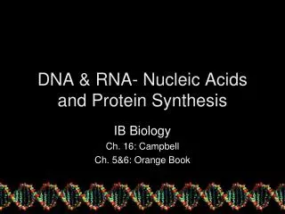
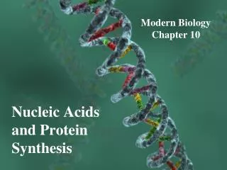
Deoxyribonucleic acid: DNA • A pair of molecules, in the form of a double helix • The 2 strands are antiparallel • Contains the genetic instructions for the development of all cellular forms of life • Wikipedia: DNA is a long polymer of nucleotides (similarly for RNA , but RNA is single stranded)
Nucleotides • 5 nucleotides: adenine (A), thymine (T), uracil (U), cytosine (C), and guanine (G). • U rarely found in DNA • T rarely found in RNA • Form complimentary pairs through hydrogen bonds: • A to T forming two bonds • C to G forming three bonds
From DNA to Proteins • The sequence of nucleotides along a DNA strand defines a messenger RNA sequence which then defines a protein • Genetic code: relation between nucleotide sequence in DNA and amino-acid sequence in proteins. • Wikipedia: The genetic code consists of three-letter 'words' (termed a codon) formed from a sequence of three nucleotides (e.g. ACT, CAG, TTT). • codons can be translated with messenger RNA and then transfer RNA, with a codon corresponding to a particular amino acid. • there are 64 possible codons (4 bases in 3 places 43) that encode 20 amino acids. Most amino acids, therefore, have more than one possible codon. • there are three 'stop' or 'nonsense' codons signifying the end of the coding region, namely the UAA, UGA and UAG codons. • Only a small fraction of the total sequence of the genome appears to encode proteins • only about 1.5% of the human genome consists of protein-coding exons . The function of the rest is a matter of speculation.
Protein molecule • Are build up by amino acids that are sequentially linked by bonds in a polypeptide chain • There are 20 amino acids, specified by genetic code • They define the primary structure (main chain, protein’s backbone) • Amino acid has: • Central carbon atom Cα attached to: • Amino group H-N-H • Carboxyl group O=C’-O-H • Hydrogen • Side chain
Protein backbone • Protein: a polypeptide chain • Amino acids on the chain: • Common part • Differ in side chains (20 possible side chains) • Define the primary structureof the protein • The repeating units are called residues • divided into • main chain atoms • side chains • main chain part is identical in all residues • Adjacent residues have peptide bonds: • Carboxyl group of residue i forms a bond, C’-N, with the amino group of residue i+1. • Consequence: the chain extends from amino terminus (first residue) to carboxyl terminus (last residue) • Biochemical viewpoint: basic repeating unit is NH—CαH—C’=O
Side chains • 20 different side chains, corresponding to 20 amino acids • Divided into 3 classes: • Hydrophobic (non-polar) • Charged (weak acid or weak base) • Polar (hydrophilic) • Amino acids (except glycine) can have two forms: L-form or D-form • Only one form, the L-form, exists in proteins • When looking along the line through H and Cα atom, the left-to-right order reads CO-R-N (yes, CORN)
Protein synthesis • Wikipedia: Within the nucleus of the cell (light blue), genes (DNA, dark blue) are transcribed into RNA. This RNA is then subject to post-transcriptional modification and control, resulting in a mature mRNA (red) that is then transported out of the nucleus and into the cytoplasm (peach), where it undergoes translation into a protein. mRNA is translated by ribosomes (purple) that match the three-base codons of the mRNA to the three-base anti-codons of the appropriate tRNA. Newly synthesized proteins (black) are often further modified, such as by binding to an effector molecule (orange), to become fully active.
Translation • Protein is grown from one end to another, by joining amino acids • Wikipedia: the correct tRNA, linked to a specific amino acid, is directed to the ribosome to be added to a growing (nascent) polypeptide • as the ribosome travels down the mRNA one codon at a time, another tRNA is attached to the mRNA at one of the ribosome sites. The first tRNA is released, but the amino acid that is attached to the first tRNA is now moved to the second tRNA, and binds to its amino acid • when the entire unit reaches the end codon on the mRNA, it falls apart and a newly formed protein is released • protein folding is done during or after synthesis • native secondary and tertiary structures can be formed during synthesis • most proteins fold to a unique, native state