Download
1 / 15
150 likes | 623 Views
Oral Health and Hygiene Training . Director: Rhonda A. Kalasho Email: rhondakalasho@gmail.com Phone number: 619729-8068 Apprentice: John Zimmerman Email: johnny.zimmerman@gmail.com. Tooth Anatomy .

E N D
Oral Health and Hygiene Training Director: Rhonda A. Kalasho Email: rhondakalasho@gmail.com Phone number: 619729-8068 Apprentice: John Zimmerman Email: johnny.zimmerman@gmail.com
Tooth Anatomy Crown— the top part of the tooth, and the only part you can normally see. The shape of the crown determines the tooth's function. For example, front teeth are sharp and chisel-shaped for cutting, while molars have flat surfaces for grinding. • Enamel— the outermost layer of the tooth. Enamel is the hardest, most mineralized tissue in the body — yet it can be damaged by decay if teeth are not cared for properly. • Dentin — the layer of the tooth under the enamel. If decay is able to progress its way through the enamel, it next attacks the dentin — where millions of tiny tubes lead directly to the dental pulp. • Pulp — the soft tissue found in the center of all teeth, where the nerve tissue and blood vessels are. If tooth decay reaches the pulp, you usually feel pain. • Root — the part of the tooth that is embedded in bone. The root makes up about two-thirds of the tooth and holds the tooth in place.
Tooth Anatomy Continued Cementum - a layer of tough, yellowish, bone-like tissue that covers the root of a tooth. It helps hold the tooth in the socket. The cementum contains the periodontal membrane. • Periodontal membrane/ligament - the fleshy tissue between tooth and the tooth socket; it holds the tooth in place. The fibers of the periodontal membrane are embedded within the cementum. • Nerves - nerves transmit signals (conveying messages like hot, cold, or pain) to and from the brain.
Teeth Numbering and Naming • Tooth number 1 is the tooth farthest back on the right side of your mouth in the upper (maxillary) jaw. • Numbering continues along your upper teeth toward the front and across to the tooth farthest back on the top left side (which is number 16). • The numbers continue by dropping down to the lower (mandibular) jaw. Number 17 is the tooth farthest back on the left side of your mouth on the bottom. • Numbering continues again toward the front and across to the tooth farthest back on the bottom right side of your mouth (which is number 32).
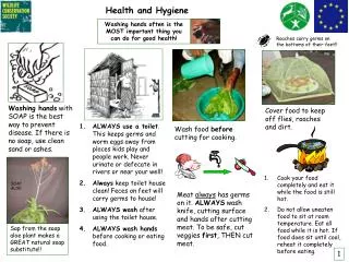
How to Brush Properly Soft bristle toothbrush Tilt toothbrush to a 45 degree angle against the gum line and gently brush in circular motion Inner tooth surface continue to brush at 45 degrees back and forth in circular motion Brush the front teeth vertically in a soft circular motion to remove any pellicle ( protein produced by saliva which house cariogenic bacteria- like streptococci which is more commonly known as plaque) Also brush biting surfaces DO NOT FORGET THE TONGUE! The tongue can house over 300 million plaque producing bacteria only after a couple of hours after brushing
An apple a day keeps the doctor away… not true unless you FLOSS ! • Cut about 18 inches off floss- about arms length • Rap around middle finger or pointer finger • Grasp • Have about ½ inch of space available to fit snuggly between teeth • Use a C-shape motion around the tooth to remove particles that found its way underneath the gum line • Use a clean section of floss each time
Dental Caries (Cavities) • The best strategies to rid your mouth of dental caries are to limit the amount of sucrose ingested, as well as brushing, flossing, and provisional dental cleanings. • Sucrose is converted by plaque causing bacteria(streptococci) into lactic acid which eats through the enamel of the tooth. • Fact: prior to the 17th century only about 10% of human remains from older times had dental caries. It was the introduction of table sugar, which is extremely cariogenic, that caused cavities to claim the mouths of tens of millions of Americans each year. • Fact: Ancient Chinese used urine as a mouthwash to help keep their teeth strong. If the Chinese knew the science behind it or not, they were nonetheless successful in neutralizing the lactic acid
Let’s play Dentist • scenario: Jane Doe walks into your practice with her hand over her lower lip. She appears to be in pain and begins to explain to you her symptoms. • Jane Doe expresses to you that tooth 23 (lateral incisor) is causing her pain as she bites down on it. It is sensitive when she eats soup and drinks hot coffee. When she eats ice-cream, it stings for more than a couple of seconds. • Jane Doe points out that her tooth is also becoming discolored. In fact, the tooth appears to be darker than all the surrounding teeth. • She also has noted that her gum line below the tooth is swollen. • Jane Doe finally mentions to you that approximately 5 months ago she chipped this tooth while chewing a Jaw Breaker. • So you take an X-ray…
What is your verdict on Jane Doe? • Keeping in mind Jane Doe’s symptoms, employ your trained eye to decipher the x-ray. How do you propose to treat Jane Doe- more specifically, with what form of treatment, and why did you come to this conclusion?
Jane Doe’s symptoms are a map to the answer- ROOT CANAL TREATMENT • Sensitivity to hot and cold- When Jane cracked her tooth this gave ample space for bacteria to spread and develop. Referencing to the x-ray, the bacteria seemed to destroy the pulp. The inflammation and infection spread down the root canal, often causing sensitivity to hot or cold foods. • Intrinsic discoloration — This is when the inner structure of the tooth (the dentin) darkens or gets a yellow tint. This occurs when there is possible root damage. • The X-ray- What you see as grey/black on x-ray: Decay, abscess, nerves and blood vessels (the pulp) and gum in the spaces between teeth. What you see as white/cream on x-ray are enamel, metal fillings and crowns will be white, and the dentine appears as a creamy white color.
Treatment • 1. Make a small hole in the tooth to reach the infected pulp. • 2. Measure the root canal to make sure all the diseased tissue is cleaned. An Apex locator may be used for this. - Dentists use X-rays to determine the length of the canals or use an electric device called an apex locator.(An apex locator makes a calculation based on the resistance to a small electric current.) 3. Use of antiseptic solution to clean the area 4. Fill the tooth and place a fitted crown
Dentist are the keepers of the ENTIRE mouth, including the tongue. • Red Tongue: If your tongue turns a shade of red you might be having a deficiency of certain nutrients. The number one cause for this is lack of niacin (B3 vitamin found in chicken breast and tuna). Another would be not getting enough folic acid in your diet or anemia. • Yellow tongue: This usually means that you have a fungus growing on your tongue. Sometimes it can be caused from having a disease called acid reflux where the acid backs up into your mouth and changes the environment there and your tongue color changes to yellow. (brushing with hydrogen peroxide can take care of the issue most of the time.
Oral Thrush or White tongue: Oral thrush is a fungal infection of the mouth, the condition where the mouth is invaded by candida albicans fungus. The organism normally lives in the mouth in controlled quantity. Cured with anti-fungal mouthwash. • Black Hairy Tongue: Black hairy tongue (lingua villosa nigra) is the excessive growth of fungi that normally live in the mouth. Bacteria can then accumulate on the finger-like projections from the surface of the tongue. The black color comes from the blood cell pigments. Treated with antifungal mouthwash and tongue scraping.
Have a Great Semester!If you have any questions about the quiz please feel free to email me at rhondakalasho@gmail.com, or call me at 6197298068. the quiz is tentatively scheduled for September 21st 2009. STUDY!