Download
1 / 8

E N D
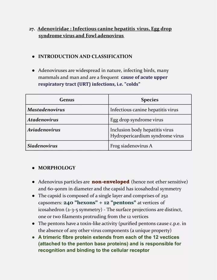
27. Adenoviridae : Infectious canine hepatitis virus, Egg drop syndrome virus and Fowl adenovirus ● INTRODUCTION AND CLASSIFICATION ● Adenoviruses are widespread in nature, infecting birds, many mammals and man and are a frequent cause of acute upper respiratory tract (URT) infections, i.e. "colds" Genus Species Mastadenovirus Infectious canine hepatitis virus Atadenovirus Egg drop syndrome virus Aviadenovirus Inclusion body hepatitis virus Hydropericardium syndrome virus Siadenovirus Frog siadenovirus A ● MORPHOLOGY ● Adenovirus particles are non-enveloped (hence not ether sensitive) and 60-90nm in diameter and the capsid has icosahedral symmetry ● The capsid is composed of a single layer and comprises of 252 capsomers: 240 "hexons" + 12 "pentons" at vertices of icosahedron (2-3-5 symmetry) - The surface projections are distinct, one or two filaments protruding from the 12 vertices ● The pentons have a toxin-like activity (purified pentons cause c.p.e. in the absence of any other virus components (a unique property) ● A trimeric fibre protein extends from each of the 12 vectices (attached to the penton base proteins) and is responsible for recognition and binding to the cellular receptor
● CULTIVATION ● The adenoviruses can be cultivated in cell cultures of natural host or·closely related species ● The CPE is characterized by rounding or clumping of affected cells resembling 'bunches of grapes' ● The CPE is produced late, 7-10 days longer ● Since the virus multiplies in nucleus, the nuclear changes include formation of basophilic or acidophilic inclusion ● The inclusions represent aggregation of proteinaceous material and crystal of mature and immature particles ● TRANSMISSION ● The virus is NOT transmitted through droplets ● The disease can be transmitted by placing urine or saliva in the oral cavity of susceptible dog - Similar to leptospirosis ● The virus is shed through urine, saliva and faeces during acute illness ● In addition to excretion, the major source of spread in birds could be the egg transmission of the virus ● PATHOGENESIS ● Following initial multiplication at the site of virus entry there is viraemia resulting in virus spread to virtually all organs ● The blood brain barrier normally prevents entry of virus into the central nervous system ● The main sites of virus replication appear to be in trachea and caeca ● Tonsils and Peyer patches get infected and cause vireamia ● The virus has got affinity for epithelial, mesothelial and hepatic cells ● The virus damages these cells causing haemorrhages and necrosis ● The liver, spleen and superficial LN are enlarged, congested, edematous and haemorrhagic
● CLINICAL SIGNS AND LESIONS a) INFECTIOUS CANINE HEPATITIS - Rubarth disease ● Clinical signs start with biphasic fever of >104°F (40°C), which lasts 1-6 days associated with leukopenia, which persists throughout the febrile period ● The degree of leukopenia varies and seems to be correlated with the severity of illness ● Other signs are apathy, anorexia, thirst, conjunctivitis, serous discharge from the eyes and nose, and occasionally abdominal pain ( due to swelling of liver capsule) and vomiting, intense hyperemia or petechiae of the oral mucosa and enlarged tonsils ● Subcutaneous edema of the head, neck, and trunk are also observed ● The blood clotting time also varies with severe haemorrhage, which is manifested by bleeding around deciduous teeth and by spontaneous hematomas ● Severely infected dog may have a terminal convulsion with brain- stem hemorrhages - On recovery, dogs will regain weight slowly ● Further, seven to 10 days after the acute signs disappear, recovered dogs develop bilateral corneal opacity, which usually disappears spontaneously - HEPATITIS BLUE EYE due to Iridocyclitis and corneal oedema ● In mild cases of ICH, transient corneal opacity may be the only sign of disease ● Chronic hepatitis develop in dogs having low levels of passive antibody when exposed ● Simultaneous infection with CAV-1 and distemper virus is also seen
● The important lesions are hepatic cell necrosis leading to change in colour of the liver, which may be normal in size or swollen ● The gallbladder wall may be edematous and thickened ● Grayish white foci are also seen in the kidney cortex ● Damage to the endothelium of intestine results in “paintbrush” hemorrhages on the gastric serosa, lymph nodes, thymus, pancreas, and subcutaneous tissues
b) EGG DROP SYNDROME ● The infection occurs in three forms classical, endemic and sporadic ● EDS affects only layers and breeders at the start of or during their egg production ● Affected flocks show a failure to reach peak egg production or a drop in egg production accompanied by an inferior eggshell quality and in the case of brown eggs, a loss of shell color ● Birds tend to eat the shell-less eggs, which therefore may be missed unless a search is made for the membranes ● Affected birds may also appear to be anaemic, may show transient diarrhoea and sometimes the food intake may be reduced ● No increased mortality or other symptoms are observed ● Birds with antibody slow the spread of virus - There is no effect on fertility or hatchability of those eggs suitable for setting ● In cage units the virus spread is slow and the clinical signs are overlooked and the problem identified as a small depression (2-4%) of egg yield ● The major pathological changes occur in the pouch shell gland ● Surface epithelial cells develop intranuclear inclusion bodies and degenerate, and are replaced by squamous, cuboidal, or undifferentiated columnar cells
c) INCLUSION BODY HEPATITIS ● The disease mainly occurs in broilers and mortality rate varies from 2-10% ● There is marked anaemia, icterus of skin, subcutaneous fat deposits, haemorrhages of various organs and pale inactive bone marrow ● At post mortem the liver is swollen, light brown to yellow with haemorrhages ● Eosinophilic inclusion bodies are present in hepatocytes ● Adenoviruses of different serotypes have been isolated from the cases of IBH ● The chicks which suffer from infectious bursal disease become immunosuppressive and lead to IBH outbreaks ● In some outbreaks the bone marrow lesions are most prominent, and it has been suggested that the disease should be called, Hepato-myeloporetic disease
d) HYDROPERICARDIUM SYNDROME - Leechi heart disease ● The affected chicks died suddenly ● The pericardial sac is filled with straw color fluid ● Swollen and discolored liver, Swollen kidneys with distended ureters ● Due to malfunctioning of liver and kidneys, there is reduction in osmotic pressure resulting in seepage of fluid in pericardial sac ● The pericardial sac becomes distended due to excessive accumulation of fluid giving rise the appearance of LICHI
● DIAGNOSIS ● Clinical signs are difficult to differentiate from canine distemper. Sudden onset with severe bleeding is suggestive of ICH ● Abnormal eggs and pouch shell glands indicates adenovirus infection ● The symptoms should be correlated with blood picture before arriving at final diagnosis ● Histopathology of liver and demonstration of specific intranuclear inclusion bodies ● Cell culture systems developed from kidney cells of dogs or ferrets are used for isolation ● The presence of virus is confirmed by demonstration of intranuclear inclusion bodies by Immunofluorescence or Polymerase chain reaction ● PREVENTION AND CONTROL ● Vaccination against ICH is recommended at the time of canine distemper vaccinations ● Attenuated CAV-1 vaccines may produce transient unilateral or bilateral opacities of the cornea, and the virus may be shed in urine. However, CAV-2 attenuated live virus strains, which provide cross protection against CAV-1 does not produce corneal opacities or uveitis, and virus is not shed in urine ● Animals should be revaccinated annually ● Inactivated vaccines with oil adjuvant available for fowl adenoviruses ● They reduce but do not prevent virus shedding - These vaccines are given during the growing phase, usually at 14- 18 wk old, and can be combined with other vaccines such as for Newcastle disease 1 1Dixit Kumar