Download
1 / 35
350 likes | 862 Views
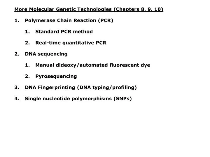
More Molecular Genetic Technologies (Chapters 8, 9, 10) Polymerase Chain Reaction (PCR) Standard PCR method Real-time quantitative PCR DNA sequencing Manual dideoxy/automated fluorescent dye Pyrosequencing DNA Fingerprinting (DNA typing/profiling)

E N D
More Molecular Genetic Technologies (Chapters 8, 9, 10) • Polymerase Chain Reaction (PCR) • Standard PCR method • Real-time quantitative PCR • DNA sequencing • Manual dideoxy/automated fluorescent dye • Pyrosequencing • DNA Fingerprinting (DNA typing/profiling) • Single nucleotide polymorphisms (SNPs)
Lots of practical applications, virtually unlimited: • Amplify DNA for Cloning (PCR) • Amplify DNA for sequencing without cloning (PCR) • DNA sequencing reaction (PCR) • Mapping genes and regulatory sequences • Linkage analysis (identify genes for traits/diseases) • Diagnose disease • Pathogen screening • Sex determination • Forensic analysis • Paternity/maternity (relatedness) • Behavioral ecology studies (relatedness) • Molecular systematics and evolution (comparing homologous sequences in different organisms) • Population genetics (theoretical and applied) • Physiological genetics (studying basis of adaptation) • Livestock pedigrees (optimize breeding) • Wildlife management (stock identification/assessment) • Detection of Genetically Modified Food (GMOs)
Polymerase Chain Reaction (PCR) • Ability to generate identical high copy number DNAs made possible in the 1970s by recombinant DNA technology (i.e., cloning). • Cloning DNA is time consuming and expensive (>>$15/sample). • Probing libraries can be like hunting for a needle in a haystack. • PCR, “discovered” in 1983 by Kary Mullis, enables the amplification (or duplication) of millions of copies of any DNA sequence with known flanking sequences. • Requires only simple, inexpensive ingredients and a couple hours. DNA template Primers (anneal to flanking sequences) DNA polymerase dNTPs Mg2+ Buffer • Can be performed by hand or in a machine called a thermal cycler. • 1993: Nobel Prize for Chemistry
How PCR works: Begins with DNA containing a sequence to be amplified and a pair of synthetic oligonucleotide primers that flank the sequence. Next, denature the DNA to single strands at 94˚C. Rapidly cool the DNA (37-65˚C) and anneal primers to complementary single-straned sequences flanking the target DNA. Extend primers at 70-75˚C using a heat-resistant DNA polymerase such as Taq polymerase derived from Thermus aquaticus. Repeat the cycle of denaturing, annealing, and extension 20-45 times to produce 1 million (220) to 35 trillion copies (245) of the target DNA. Extend the primers at 70-75˚C once more to allow incomplete extension products in the reaction mixture to extend completely. Cool to 4˚C and store or use amplified PCR product for analysis.
Hot water bacteria: Thermus aquaticus Taq DNA polymerase Life at High Temperatures by Thomas D. Brock Biotechnology in Yellowstone © 1994 Yellowstone Association for Natural Science http://www.bact.wisc.edu/Bact303/b27
Fig. 9.3 Denature Anneal PCR Primers Extend PCR Primers w/Taq Repeat…
Example thermal cycler protocol used in lab: Step 1 7 min at 94˚C Initial Denature Step 2 45 cycles of: 20 sec at 94˚C Denature 20 sec at 52˚C Anneal 1 min at 72˚C Extension Step 3 7 min at 72˚C Final Extension Step 4 Infinite hold at 4˚C Storage
Real-time Quantitative PCR: Measures the abundance of DNA as it is amplified. Useful for quantitatively measuring the levels of mRNA in a sample. Uses reverse transcriptase to generate cDNA for the template. Can also be used to quantitatively estimate fraction of DNA from various organisms in a heterogenous sample (e.g, can be used to measure abundance of different microbes in soil sample). Fluorescent dye, SYBR Green, is incorporated into PCR reaction. SYBR Green fluoresces strongly when bound to DNA, but emits little fluorescence when not bound to DNA. SYBR Green fluorescence is proportional to the amount of DNA amplified, detected with a laser or other device. Experimental samples are compared to control sample with known concentration of cDNA.
Fig. 10.9 SYBR Green binds to double-stranded DNA and fluoresces
DNA Sequencing • DNA sequencing = determining the nucleotide sequence of DNA. • Dideoxy sequencing developed by Frederick Sanger in the 1970s. 1980: Walter Gilbert (Biol. Labs) & Frederick Sanger (MRC Labs)
Dideoxy DNA sequencing relies on chain termination: • DNA template is denatured to single strands. • DNA primer (with 3’ end near sequence of interest) is annealed to the template DNA and extended with DNA polymerase. • Four reactions are set up, each containing: • DNA template • Primer annealed to template DNA • DNA polymerase • dNTPS (dATP, dTTP, dCTP, and dGTP) • Next, a different radio-labeled dideoxynucleotide (ddATP, ddTTP, ddCTP, or ddGTP) is added to each of the four reaction tubes at 1/100th the concentration of normal dNTPs. • ddNTPs possess a 3’-H instead of 3’-OH, compete in the reaction with normal dNTPS, and produce no phosphodiester bond. • Whenever the radio-labeled ddNTPs are incorporated in the chain, DNA synthesis terminates. • Each of the four reaction mixtures produces a population of DNA molecules with DNA chains terminating at all possible positions.
Dideoxy DNA sequencing (cont.): Extension products in each of the four reaction mixtures also end with a different radio-labeled ddNTP (depending on the base). Next, each reaction mixture is electrophoresed in a separate lane (4 lanes) at high voltage on a polyacrylamide gel. Pattern of bands in each of the four lanes is visualized on X-ray film. Location of “bands” in each of the four lanes indicate the size of the fragment terminating with a respective radio-labeled ddNTP. DNA sequence is deduced from the pattern of bands in the 4 lanes.
Fig. 8.17, 2nd edition Vigilant et al. 1989 PNAS 86:9350-9354
Radio-labeled ddNTPs (4 rxns) Sequence (5’ to 3’) G G A T A T A A C C C C T G T Short products Long products
Automated Dye-Terminator dideoxy DNA Sequencing: Original dideoxy DNA sequencing methods were time consuming, radioactive, and throughput was low, typically ~300 bp per run. Automated DNA sequencing employs the same general procedure, but uses ddNTPs labeled with fluorescent dyes. Combine 4 dyes in one reaction tube and electrophores in one lane on a capillary containing polyacrylamide. UV laser detects dyes and reads the sequence. Sequence data is displayed as colored peaks (chromatograms) that correspond to the position of each nucleotide in the sequence. Throughput is high, up to 1,200 bp per reaction and 96 reactions every 3 hours with capillary sequencers. Most automated DNA sequencers can load robotically and operate around the clock for weeks with minimal labor.
Applied Biosystems PRISM 377 (Gel, 34-96 lanes) Applied Biosystems PRISM 3700 (Capillary, 96 capillaries) Applied Biosystems PRISM 3100 (Capillary, 16 capillaries)
“virtual autorad” - real-time DNA sequence output from ABI 377 Trace files (dye signals) are analyzed and bases called to create chromatograms. Chromatograms from opposite strands are reconciled with software to create double-stranded sequence data.
Pyrosequencing: • Based on the “sequencing by synthesis” principle instead of chain termination with dideoxy nucleotides. • Developed by Pål Nyrén/Mostafa Ronaghi in 1996. • Immobilize a single template DNA molecule on a bead and synthesize complementary strand. • Detect which nucleotide is added at each step. • Requires template DNA, primer, DNA polymerase, ATP sulfurylase, luciferase, apyrase, adenosine 5’ phosphosulfate (APS), and luciferin.
Fig. 8.12 Example showing how to read pyrosequencing data
Maximum pyrosequencing read lengths currently are 300-500 nt. Commercial applications: 454 Life Sciences Genome Sequencer FLX Generate 400 million nt in 10 hours $5-7K USD per run $1M for mammalian genome
DNA Fingerprinting (DNA typing/profiling) • No two individuals produced by sexually reproducing organisms (except identical twins) have exactly the same genotype. Why? • Crossing-over of chromosomes in meiosis prophase I. • Random alignment of maternal/paternal chromosomes in meiosis metaphase I. • Mutation • DNA replication errors (same effect as mutation)
DNA Fingerprinting (DNA typing/profiling) Types of markers: • RFLPs (restriction sites) • Length polymorphism detected by PCR • Allele specific oligonucleotide probes • Repeated DNA • Minisatellites (VNTRs = variable number tandem repeats) Repeated units of 5 to several 10 bp Discovered by A. J. Jeffreys in 1985 • Microsatellites (STRs = short tandem repeats) Repeated units of 2-6 bp 5’-TAATAATAATAATAATAA-3’ 3’-ATTATTATTATTATTATT-5’
Four criteria for selecting useful DNA fingerprinting markers: • Markers should be polymorphic. (so that they are informative) • Markers should be single locus. (so that they occur in only one location in the genome and there is no ambiguity about their number or position) • Markers should be located on different chromosomes. (so that the markers are independent) • For some applications such as the study of population size and changes over time, markers should be neutral. (so that they are not correlated with selection or adaptation; unless selection of adaptation are to be studied; selection confounds estimates of population size parameters)
Microsatellites (short tandem repeats or STRs): Heterozygote Male 5’-TAATAATAATAATAATAATAA----3’ Female 5’-TAATAATAATAATAATAATAATAA-3’ Homozygote Male 5’-TAATAATAATAATAA-3’ (different allele) Female 5’-TAATAATAATAATAA-3’ • One proposed explanation for their fast rate of evolution is slippage during DNA replication. • Excellent marker for DNA fingerprinting because: • Polymorphic (fast-evolving) • Single locus • Common throughout genomes of most organisms • Neutral (non-coding)
How to fingerprint alleged paternity using microsatellites: Extract DNA from mother, baby, and alleged father. Synthesize oligonucleotide microsatellite primers and label one primer with fluorescent dye (2 primers per microsatellite). Amplify microsatellites using PCR from mother, baby, father. Electrophores microsatellite PCR products on a DNA sequencer (w/polyacrylamide) with a flourescent size standard loaded in the same lane or capillary. 3-4 different microsatellites can be multiplexed in each lane or capillary by using 3-4 different fluorescent dyes. Calculate size of each microsatellite relative to size standard (this size standard also can be run in the same gel lane or capillary using a 4th or 5th colored dye). Sequence at least one copy of each allele to verify allele sizes.
Size Mother Baby “Father” Hypothetical gel pattern for microsatellite heterozygous for all individuals.
Paternity Analyses & Conclusions: • Baby and mother are expected to share on allele, and the baby and father the other allele. • If baby and father do not share a common allele, the “father” is not the father. • If the baby and father do share a common allele, paternity is possible, but not proven, because other men in the population also carry the allele at some frequency. • Frequency of alleles that are shared in common by chance can be calculated, and an appropriate number of microsatellites analyzed to calculate probability of paternity. • To achieve high probability, 6-12 loci should be assayed (exact number depends on variation in population for each marker). • If each locus has few alleles, more loci are required. If allelic diversity if high, fewer loci can be analyzed.
Single nucleotide polymorphisms (SNPs): DNA sequences of most individuals are almost identical, >99%. Single base pair differences occur about once every 500-1000 bp. In most populations there is a common SNP, and several less common SNPs. About 3 million SNPs occur in the human genome, and these are becoming popular genetic markers. SNPs can be used just like other genotyping markers, but more loci typically must be used because only 4 alleles (G, G, C, T) are possible.
How to type SNPs: • SNPs can be typed by hybridizing a complementary oligonucleotide (e.g., single-base extension assay). • If the stringency is high (i.e., temperature), the oligonucleotide will fail to bind to DNAs showing polymorphism. • Many hundreds of SNPs can be tested simultaneously using: DNA microarrays (DNA-chips, Gene-Chips, SNP-chips) • First developed in the early 1990s. • Ordered grid of short, complementary, known sequence oligonucleotides placed at fixed positions on silicon, glass, or nylon substrate. • Oligonucleotides are experimentally determined and are either (1) microspotted or (2) synthesized on the chip. • User defined SNP chips are available commercially, and can contain >400,000 different probes.
How to type SNPs (cont.): SNP chip is designed with an array of user defined oligonucleotides attached to the substrate (the SNP chip is the probe). Oligonucleotides match each of the common and variant alleles in the population (all alleles of interest). Target DNAs are labeled with a fluorescent tag and hybridized (or not) to the chip. Fluorescence pattern is detected by a laser. Because the oligonucleotides are known, the pattern indicates the type of alleles the individual possesses. Many different alleles at thousands of different loci can be screened simultaneously in the same experiment.