Download
1 / 58
1.42k likes | 6.8k Views
Extracorporeal Membrane Oxygenation (ECMO). Dr. Yan Wing Wa, MBBS, MSc, MRCP, FRCP(Lond, Edin), FHKCP, FHKAM(Medicine) Chairman, Specialty Board in Critical Care Medicine, Hong Kong College of Physicians President, Hong Kong Society of Critical Care Medicine

E N D
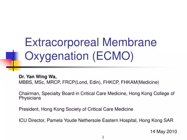
Extracorporeal Membrane Oxygenation (ECMO) Dr. Yan Wing Wa, MBBS, MSc, MRCP, FRCP(Lond, Edin), FHKCP, FHKAM(Medicine) Chairman, Specialty Board in Critical Care Medicine, Hong Kong College of Physicians President, Hong Kong Society of Critical Care Medicine ICU Director, Pamela Youde Nethersole Eastern Hospital, Hong Kong SAR 14 May 2010
ECMO • A form of extracorporeal life support where an external artificial circuit carries venous blood from the patient to a gas exchange device (oxygenator) where blood becomes enriched with oxygen and has carbon dioxide removed. This blood then re-enters the patient circulation. • Flow ~70ml/kg/min • ~3ml/kg/min in CRRT
Evolution of ECMO • Robert H Bartlett, MD • Director of the Extracorporeal Life Support Program • The University of Michigan Extracorporeal Life Support Team • Largest ECMO experience in the world (>1,000 cases prior to 2000) • 1985 Prospective Randomised Trial in Neonatal Respiratory Failure, Pediatrics 1985,76(4)479-87 • 1 patient in conventional arm (died) • 11 patients in the study arm (all survived)
CESAR studyConventional ventilation or ECMO for Severe Adult Respiratory failureLancet 2009, 374:1351-63 • Survival without severe disability (confined to bed, or unable to dress/wash oneself) by 6 months • ECMO: 57 in 90 patients (63%) • Conventional ventilation: 41 in 87 patients (47%) • Relative risk reduction in favour of ECMO 0.69 (0.05–0.97; P = 0.03) • NNT to prevent one death is 6
Veno-venous (VV) ECMO R L R L
VV-ECMOAdvantages & disadvantages • Advantages • Normal lung blood flow • Oxygenated lung blood • Pulsatile Blood Pressure • Oxygenated blood delivered to root of aorta • Must be used when native cardiac output is high • Disadvantages • No Cardiac support • Local recirculation through oxygenator at high flows • Reversed gas exchange in lung if FiO2 low • Limited power to create high oxygen tensions in blood
VV-ECMO • Single drainage cannula • Efficient CO2 removal • Weak effect on Oxygenation • Use for respiratory indications when severe hypoxia is not a problem
VV-ECMO (Hi-flow) • Twodrainage cannulae • Effectivenessof high flow limited by recirculation from return to drainage cannulae • Oxygenationlimited by effective flow (total-recirculated) (but not a problem for CO2) • Usedin lung conditions with severe hypoxia
VA-ECMO • Central (ascending aorta) • During sternotomy/ via subclavian artery • VA-ECMO for CPR • Simple and rapid to establish • Temporary for retrieval • Limb ischaemia • Hi blood flow VA-ECMO • Double drainage cannulae • Distal limb perfusion
VA-ECMO for CPR Hi Blood flow VA-ECMO
VA-ECMOAdvantages & disadvantages • Disadvantages: NO • Normal lung blood flow • Oxygenated lung blood • Pulsatile Blood Pressure • Oxygenated blood delivered to root of aorta (except central) • Advantages • Cardiac support also • No local recirculation through oxygenator at high flows • No reversed gas exchange in lung • Power to create high oxygen tensions in blood
Quadrox® PLS oxygenator • Low pressure drop • Efficient integrated heat exchanger • CE certified continous use for 14 days • Low priming volume 250ml • Low membrane surface area 1.8m2 • Very high transfer rate of O2 and CO2
Jostra Centrifugal Pump • Jostra RotaFlow impeller pump • 32ml priming volume • The RotaFlow had no stagnant blood zones, no shaft and no seals
Objectives • H1N1 pandemic in 2009 • After a review of the published literatures • CESAR study • Australian & New Zealand H1N1 ECMO study • Introduce veno-venous extracorporeal membrane oxygenation (VV-ECMO) to the Intensive Care Unit (ICU) as a rescue therapy for potentially reversible refractory hypoxaemic patients.
Scopes • For ICU medical and nursing staff. • Since ECMO service is still in its early stage of development in Hong Kong, changes will likely be made to this document with accumulation of experience in concordance with the Capability Maturity Model.
Indications for VV-ECMO • Potentially reversible and life-threatening respiratory failure unresponsive to optimum conventional ventilation and therapy. • Severe respiratory failure was defined in the CESAR trial as: • Murray score* ≥3; or • Uncompensated hypercapnia with pH ≤ 7.20
Absolute contraindications • Advanced malignancy or any fatal diagnosis • Unwitnessed cardiac arrest • Progressive and non-recoverable respiratory disease • Severe pulmonary hypertension and right ventricular failure (mean PAP approaching systemic pressure) • Severe cardiac failure: consideration should be given to veno-arterial (VA)-ECMO • Immunosuppression • Transplant recipients beyond 30 days • Advanced HIV defined by secondary malignancy, prior hepatic or renal failure (cirrhosis or serum creatinine >250μmol/L), or requiring salvage anti-retroviral treatment • Recent diagnosis of haematological malignancy • Bone marrow transplant recipients • Body size <20kg or >120kg
Relative contraindications • Preexisting conditions which affect the quality of life • Age >70 year-old • CPR duration >60 minutes • Multiple organ failure • Central nervous system injury • Contraindication to anticoagulation (no citrate) • Patient who had been on high pressure (peak pressure >30cmH20) or high FiO2 (>0.8) ventilation for >7days
Prime the circuit • Check for leakage of the heat exchanger by flushing it with water before priming the oxygenator. • The circuit is primed with normal saline (1L bag) under sterile conditions. • Make sure no bubbles in the circuit tubing, oxygenator and Rotaflow • If concomitant CVVH is required, leave behind one of the 3-way stopcocks on the venous line for connection to the dialysis machine. • The fluid in the circuit is warmed by the heat exchanger before it is attached to the patient • For HSI patients, keep >37oC
Vascular access in VV-ECMO • Select appropriately sized cannulae to provide the desired extracorporeal blood flow • The flow through a single Maquet HLS cannula at pressure drop of 60mmHg is as follows:
Vascular access in VV-ECMO (2) • If the desired blood flow cannot be achieved with a single access cannula, insert a second access cannula. • Decide on 2 cannulation sites for blood drainage and return. • Jugular vein cannulation is contraindicated in unilateral internal jugular vein thrombosis. • Cannulation into the subclavian vein for ECMO is not preformed. • Xray, fluro or echocardiogram can be used to guide cannula positioning. • The access and return cannulae should be placed at some distance apart to minimize access recirculation.
The cannulation sites are dressed and covered with Tegaderm. The cannula and tubing are firmly secured to the skin with non-circumferential Elastoplast or Mefix
For internal jugular vein insertion, the cannula and tubing are bound to the head with elastic bandages
Oxygenation • Begin extracorporeal blood flow at 70ml/kg/min for adults. • Titrate blood flow to maintain systemic arterial oxygen saturation while on low ventilator settings. • A systemic arterial saturation around 80% will be adequate for systemic oxygen delivery if the haematocrit is over 40% and cardiac function is good. • The absence of persistent metabolic acidosis is indicative of an adequate systemic oxygen delivery. • In-line venous saturation monitor may not reflect the true venous saturation in the presence of circuit recirculation. • If oxygenation cannot be maintained with persistent metabolic acidosis, the followings can be considered: • Increase extracorporeal blood flow • In access insufficiency, increase intravascular volume, or insert a second access cannula. • Blood transfusion to maintain a haematocrit level between 40-45% • Increase ventilator FiO2 and ventilatory support • In cardiac failure, increase cardiac output using volume, inotropes, or conversion to VA-ECMO for cardiac support.
CO2 removal • Use 100% oxygen as sweep gas. • Begin with a sweep gas flow rate of 6L/min. After the extracorporeal blood flow has been adjusted, set the sweep gas to extracorporeal blood flow ratio to 1:1 • ? A higher PaCO2 is beneficial to subsequent weaning • Titrate sweep gas flow rate according to carbon dioxide partial pressure: Increase sweep gas flow rate to increase carbon dioxide clearance
Anticoagulation • Bolus heparin 50-100 units/kg after successful cannulation followed by continuous infusion. • Continuous heparin infusion at 10-15units/kg/hour. • Titrate dose to maintain APTT of 50-60s. • A higher APTT level should be targeted for extracorporeal blood flow in the range of 0.5 to 2.5L/min. • Monitor APTT every 6 hours. Some centres may choose to monitor ACT instead of APTT
Ventilator management • While on VV-ECMO, the ventilator should be adjusted to a low setting to allow for lung rest: • Low FiO2 (<40%) • Low tidal volume (<6ml/kg ideal body weight) and peak airway pressure (<35cmH2O) to avoid volutrauma • A higher PEEP (10-20cmH2O) to keep alveloli open and prevent atelectotrauma.
Sedation • should be thoroughly sedated at the time of cannulation and for the first 12 to 24 hours • facilitate successful cannulation • avoid air embolism in the presence of spontaneous breathing • minimize metabolic rate • enhance comfort. • Once the patient is stable on VV-ECMO, sedation should be minimized
Possible complications • Haemolysis • Intravascular haemolysis can result from Access insufficiency: • Insufficient venous return • Obstructed access cannula • Access cannulatoo small • Clots within the circuit • Inappropriate pump speed • Bleeding • Apply direct pressure to accessible sites. • In case of bleeding at the cannulation site, rule out decannulation. • Circuit rupture • Cleaning circuit (polycarbonate components) with alcohol predisposes to fracture and should be avoided
Possible complications (2) • Pump failure • Causes: • Pump head disengagement from accidental contact or incorrect placement • Motor failure • Battery failure in the absence of AC power • Air in circuit • To prevent air embolism, it is necessary to maintain the pressure at the blood side higher than that at the gas side: • Keep the oxygenator below the level of the patient. • Clotting in circuit • Clots larger than 5mm or enlarging clots on the return side of the circuit should be removed. • Decannulation • Accidental removal of either or both cannulae.
Connections for continuous renal replacement therapy (CRRT) • For CRRT circuit, the blood drainage side is conventionally labeled as arterial, and the blood return side as venous. This is in opposite to that of the ECMO circuit. • The return line of the CRRT circuit is connected to the luer lock connector on the arterial cannula or a distal connector placed between the oxygenator and return cannula on the ECMO circuit via a 3-way tap
Connections for continuous renal replacement therapy (2) • If a Prismaflex dialysis machine with adjustable access pressure alarm or its equivalent is used, the access line of the CRRT circuit is connected to a proximal connector between the oxygenator and return cannula on the ECMO circuit via a 3-way tap. • If a dialysis machine with no adjustable access pressure alarm is used, the access line of the CRRT circuit is connected to a connector placed before the pump on the ECMO circuit via a 3-way tap
Weaning off VV-ECMO • Increase ventilator support to a setting acceptable off VV-ECMO. • Turn off the sweep gas but continue pump rate to maintain extracorporeal blood flow. • Monitor systemic arterial oxygen saturation and pCO2. If parameters remain adequate after one hour of ventilation at an acceptable setting with the sweep gas turned off, the patient is ready to come off VV-ECMO. • Stop heparin infusion once the decision has been made to come off VV-ECMO. The circuit can be removed after 4 hours
Decannulation • Involved staff should put on PPE for standard precaution. • Turn off pump and clamp lines on both the access and return sides. • Remove the cannulae. Apply direct pressure manually or with a C-clamp