Download
1 / 53
630 likes | 2.37k Views
Dent 633 Dental Amalgam. William A. Brantley (PhD) Professor and Director Graduate Program in Dental Materials Science Section of Restorative and Prosthetic Dentistry College of Dentistry, The Ohio State University Postle Hall Room 3005-L E-mail: brantley.1@osu.edu. Textbook References.

E N D
Dent 633Dental Amalgam William A. Brantley (PhD) Professor and Director Graduate Program in Dental Materials Science Section of Restorative and Prosthetic Dentistry College of Dentistry, The Ohio State University Postle Hall Room 3005-L E-mail: brantley.1@osu.edu
Textbook References O’Brien WJ (editor). Dental Materials and Their Selection (3rd ed). Chicago: Quintessence, 2002. Chapter 12. Powers JM, Sakaguchi RL (editors). Craig’s Restorative Dental Materials (12th ed). Mosby, 2006. Chapter 11. Anusavice, KJ (editor). Phillip’s Science of Dental Materials (11th ed). Saunders/Elsevier, 2003. Chapter 17.
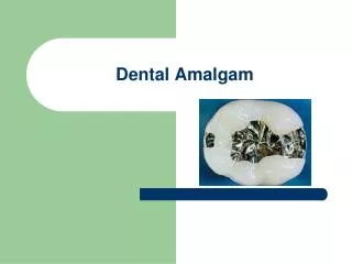
Advantages of Dental Amalgam as Restorative Material Relatively inexpensive compared to gold alloy Easily prepared direct restorative material Margin-sealing capability (decreased marginal microleakage with time) – corrosion products Over 100 years of successful clinical history (dating from GV Black dental amalgam)
Concerns about Dental Amalgam as Restorative Material Poor esthetics compared to resin composites Weakening of tooth from removal of tooth structure Recurrent caries No adhesive bonding unless bonded restoration Sensitivity of properties to manipulation Brittle nature of material Biocompatibility – not generally considered problem for patients (supported by recent JAMA article) Wastewater pollution with mercury
General Setting Reaction for Dental Amalgam Alloy (for dental amalgam) + Hg Dental amalgam Components in two compartments of capsule Mercury/alloy ratio - approximately 0.5 and depends upon particular commercial product Modern precapsulated products contain approximately 42 to 45% Hg by weight Factors for setting process: composition, shape and size of alloy particles (based on handling characteristics desired by manufacturer)
Alloy for Dental Amalgam(Particles Mixed with Mercury) ANSI/ADA specification no. 1 does not require specific percentages of elements Major element is Ag, Sn has second-largest amount, Cu about 2% to nearly 30%, Zn from 0 to about 1% Other elements if manufacturer submits results of clinical and biological testing (e.g., In and Pd) Particles have complex structure with three phases – γ (Ag3Sn), β (Ag-Sn) and ε (Cu3Sn)
Classification of Products by Particle Shape and Composition Filing or lathe-cut (machined from cast ingot) Spherical (molten alloy blown through nozzle) All particles with same composition Blend or admixture of particles with different compositions Spherical particles range from 50 μm diameter to over order of magnitude smaller — also wide range in sizes of lathe-cut particles Intentionally done for optimum condensation
Importance of Particle ShapeSpherical vs. Lathe-Cut Products Spherical particles are wetted with lower mercury:alloy ratio than lathe-cut particles Spherical particles resist forces of condensation less than lathe-cut particles
Microstructures of Lathe-Cut (100), Spherical (300), and Admixed (500) Alloys for Dental AmalgamFigures from Anusavice (11th ed), Chapter 17
Classification of Products by Alloy Composition High-copper vs. low-copper – high-copper products contain >12 % Cu in alloy particles High-copper products should be selected – greater clinical longevity of restorations and much lower creep values measured in laboratory Zinc-containing vs. zinc-free (< 0.01 wt % Zn) – not economically feasible to eliminate Zn Zinc considered to facilitate machining lathe-cut particles (more brittle) and improves corrosion resistance of amalgam, but less plastic mix No concern with Zn-free alloys about moisture contamination during trituration or condensation
Frequent Letter Codes for Dental Amalgam Products in Books and Articles LCL, LCS (low-copper alloy, lathe-cut or spherical particles) HCSS (high-copper alloy, spherical particles of single composition) HCB (high-copper alloy, blend of two different types of particles — shape and/or composition)
Composition Details for Two ProductsValiant PhD (*) –formerly used in CollegePermite C (**) –currently used in College
Heat Treatment of Alloy for Dental Amalgam by Manufacturer Eliminates compositional nonuniformity that exists in ingot before lathe-cutting (machining) or in spherical alloy particles – due to rapid freezing Relieves stresses in alloy particles (both lathe-cut and spherical) Provide manufacturer control of setting time – great clinical importance
General Form of Setting Reaction γ (starting alloy particles) + Hg (liquid) reaction phases (matrix) + unreacted alloy particles (core) Incompletely consumed alloy particles in set dental amalgam microstructure “Bricks” (alloy particles) and “mortar” (reaction phases) analogy for structure and strength of set amalgam No free mercury after setting reaction – Hg found in reaction phases Microstructure will contain some porosity from incomplete condensation
Setting Reaction ProductsThree Types of Dental Amalgams Low-copper dental amalgams – γ1 (Ag2Hg3) and γ2 (Sn8Hg) High-copper dental amalgams –γ1 and η (Cu6Sn5) Note that high-copper dental amalgams are γ2-free
Setting Reaction for Dispersalloy-Type Dental Amalgams Alloy particles are admixture or blend of low-copper lathe-cut particles and spherical Ag-Cu particles (72 wt% Ag, 28 wt % Cu) First step of setting reaction identical to low-copper dental amalgams Second step of setting reaction is disappearance of γ2 phase and formation of η phase Slower setting reaction than for HCSS products
Dimensional Changes During Setting of Dental Amalgams Total dimensional change after 24 hr < 20 μm/cm (±0.20%) is ANSI/ADA Specification no. 1 limit and cannot detect by unaided eye or explorer Most modern dental amalgam products undergo an overall contraction of the setting mass Clinical problems would occur with excessive setting expansion (loss of anatomy and postoperative pain) or excessive setting contraction (microleakage)
Setting Dimensional ChangesA: HCB, B: HCSS and C: LCLFromAnusavice (11th ed), Fig. 17-10
Nature of Setting Dimensional Changes Setting process is combination of solution and crystallization (precipitation) Initial contraction from absorption of Hg (diffusion) by amalgam alloy particles Subsequent formation and growth of γ1, γ2 and Cu-Sn phases (matrix) Final absorption of mercury by remaining amalgam alloy particles No free mercury in final set dental amalgam
Microstructure of LCL Dental AmalgamOriginal Magnification 1000From Anusavice (11th ed), Fig. 17-5
Microstructure of HCB Dental AmalgamOriginal Magnification 1000Mercury-rich droplets from polishing specimenFrom Anusavice (11th ed), Fig. 17-7
Microstructure of HCSS Dental AmalgamRelief polish with original magnification 560From Anusavice (11th ed), Fig. 17-8
Characteristics of Microstructural Phases in Dental Amalgams Strongest phase – incompletely consumed starting alloy particles (γ) Weakest phase – γ2 in low-copper amalgams (most corrosion prone) Completely interconnected nature of γ2 can result in bulk corrosion of low-copper dental amalgam High-copper amalgams – Cu6Sn5 (η) is corroding phase that provide margin-sealing – because η is not interconnected,corrosion limited to marginal regions without bulk corrosion
Types of Corrosion in Dental Amalgams Galvanic corrosion at interproximal contacts with gold alloys Electrochemical corrosion because multiple phases Crevice corrosion at margins At unpolished scratches or secondary anatomy — lower pH and oxygen concentration of saliva Corrosion under retained plaque because of lower oxygen concentration Chemical corrosion from reaction with sulfide ions at occlusal surface
Corrosion of Dental Amalgam Restorations Limited corrosion is beneficial because reduction in microleakage – γ2 in low-copper amalgams and Cu6Sn5 (η) in high-copper amalgams Tin-containing and copper-containing phases have been identified as corrosion products Corrosion minimized by polishing amalgam restoration – scratches and pits trap debris, enhancing corrosion because lower oxygen concentration under deposit Clinical trials suggest that Zn-containing amalgam restorations have superior marginal integrity and longevity – preferential Zn corrosion may occur
Mechanical Properties of Dental Amalgams Brittle material for normal rates of loading (CS » TS) and need dentinal support to resist forces of mastication Poor edge strength – fracture of ledge on poorly finished restoration readily occurs (low tensile strength leads to fracture in bending) Insufficient strength of set dental amalgam would also increase amount of marginal breakdown
Change in Mechanical Properties of Dental Amalgams with Time ANSI/ADA specification no. 1 requires specific compressive strength after 1 hr – has practical significance Rate of strength increase is dependent upon particular product ― HCSS has most rapid setting reaction Much greater difference in strength for wide range of products after 1 hr compared to 1 day Final strength considered after 1 week – nearly same strength after 1 day Mechanical properties measured in laboratory are dependent upon rate of loading – creep at constant load)
Typical Strength for Dental Amalgams (24 hr) Compressive strength > about 350 MPa Tensile strength < about 70 MPa High ratio for CS divided by TS ― brittle material
Laboratory Creep Test forDental Amalgams Cylindrical specimen stored 1 wk (37º C), compress at 36 MPa (37º C), measure length change for 1 - 4 hr Maximum creep limit in ANSI/ADA specification no. 1 High-copper amalgams generally have low creep (<1%) Creep is only mechanical property correlated with clinical marginal fracture of low-copper amalgam restorations (not high-copper which have low creep) Creep mechanism is grain boundary sliding of γ1 phase (blocked by η in high-copper amalgams)
Values of Strength and CreepRepresentative Dental AmalgamsAnusavice (11th ed), Table 17-2
Effects of Manipulative VariablesSetting Expansion of Dental Amalgams(Increased with more setting reaction phases) Excessive mercury content – increases SE Increased tritutation time – decreases SE Increased condensation pressure – decreases SE Moisture contamination of Zn-containing amalgam causes delayed, excessive increase in SE ― reason why Zn-free products often selected
Delayed Expansion of Moisture-Contaminated Zinc-Containing AmalgamFrom Anusavice (11th ed), Fig. 17-11
Effects of Manipulative VariablesStrength of Dental Amalgams(Increased with less setting reaction phases) Excessive mercury content – decreases strength Increased tritutation time – increases strength Increased condensation pressure – increases strength Moisture contamination of Zn-containing dental amalgam – large decrease in strength Zinc reduction of H2O releases H2 gas, causing excessive delayed expansion and possible postoperative pain from pulpal pressure
Effect of Mercury/Alloy RatioStrength of Dental AmalgamsFrom Anusavice (11th ed), Fig. 17-11
Some Clinical Considerations for Trituration Role of trituration ─ coat each alloy particle with mercury Overtrituration makes mixed material hot, reduces working time, and increases creep Optimum trituration time is highly important Also important to avoid undertrituration
Roles of Condensation Adapt restoration to cavity walls Minimize porosity in restoration Control final mercury content of restoration Do not delay condensation after trituration
Mercury and Mercury Toxicity Mercury is liquid metal (temperatures greater than –39°C) with high density (13.6 gm/cm3) and high vapor pressure that rapidly increases with temperature Because of mercury toxicity, US government has set threshold limit value (TLV) for sustained (40 hr/wk) exposure at 0.05 mg Hg/m3 Routes for mercury exposure - skin contact, inhalation of vapor, airborne droplets At level of 100 ng Hg per mL blood, symptoms of mercury poisoning are typically observed Some patients may exhibit an allergic skin reaction to dental amalgams
Mercury Hygiene Recommendations by ADA Use single-use capsules when preparing dental amalgams Use a no-touch technique and clean up any spilled mercury Discard any old or damaged mixing capsules which might be prone to leakage Store dental amalgam scrap in cool space in capped, unbreakable jar holding water with finely divided sulfur Avoid baseboard heating in operatories where dental amalgam is used
Mercury Hygiene Recommendations by ADA Use face mask and water spray with high vacuum evacuation when finishing new dental amalgam restorations or removing old restorations Do not use ultrasonic condensers for dental amalgam restorations Mercury vapor levels in offices and operatories where dental amalgam restorations are prepared and placed should be regularly checked Office personnel involved with dental amalgam restorations should have their mercury levels periodically monitored by urinalysis
Biocompatibility of Dental Amalgams Concern about mercury poisoning arises from high vapor pressure of liquid Hg (1.20 x 10⁻³ Torr at 20ºC), which rapidly increases with temperature Modern analytical equipment can detect mercury vapor levels as low as 1 g/m3 in air or 0.2 ng/mL in solution Possible toxicity effects from minute amounts of mercury released by dental amalgams can now be more readily investigated than previously
Biocompatibility of Dental Amalgams A clinical study (Reinhardt et al, J Prosthet Dent 1983;49:652-656) found that the amount of mercury exhaled by a patient following removal or placement of a single amalgam restoration is very small, occurs for a relatively brief period of time, and is largely avoidable by proper clinical procedures (e.g., use of rubber dam, handpiece water spray and high-volume evacuation coolant)
Biocompatibility of Dental Amalgams Theoretical calculations (Mackert, J Dent Res 1987;66:1775-1780) have shown that, even for an individual with twelve or more occlusal surfaces containing amalgam restorations and not occupationally exposed to mercury, the daily dose of mercury from these restorations would be only 10% of the normal daily intake from food, air and water
Biocompatibility of Dental Amalgams In a clinical study on patients with controlled diets (Berglund, J Dent Res 1990;69:1646-1651), the estimated average daily dose of mercury vapor inhaled from dental amalgam restorations was 1.7 g, with a range of 0.4 ‑ 4.4 g This is about 1% of the dose that would be obtained from the threshold limit value (TLV) of 50 g/m3 of airborne mercury set by the US government for an individual exposed eight hours per day for a five-day work week
Biocompatibility of Dental Amalgams Snapp et al (J Dent Res 1989;68:780-785) examined ten adults with average of fourteen restoration surfaces Mean baseline total blood Hg of 2.18 ng/mL was significantly correlated with number of occlusal surfaces After removal of amalgam restorations, nine subjects had significant decrease in blood mercury (mean 1.13 ng/mL) Removal of restorations caused exposure 1.46 ng Hg/mL, which disappeared within three days Mercury toxicity in most sensitive adults occurred at blood concentrations of 30 ng/mL, indicating that dental amalgam restorations do not appear to be health hazard
Biocompatibility of Dental Amalgams Wataha et al (Dent Mater 1994;10:298-303) placed specimens of two dental amalgams in contact with Balb/c mouse fibroblasts for 24 hr and investigated succinic dehydrogenase activity of cells as monitor of cytotoxicity HCSS dental amalgam Tytin exhibited no cytotoxicity compared to teflon controls HCB dental amalgam Dispersalloy was severely cytotoxic initially when release of Zn ions was greatest, but less toxic between 48 and 72 hr as Zn release decreased
Contrary ViewpointBiocompatibility of Dental Amalgams Lorscheider F, Vimy M. Mercury and idiopathic dilated cardiomyopathy. J Am Coll Cardiol 2000;35:819-820. Comment on J Am Coll Cardiol 1999;33:1578-1583. Aschner M, Lorscheider FL, Cowan KS, Conklin DR, Vimy MJ, Lash LH. Metallothionein induction in fetal rat brain and neonatal primary astrocyte cultures by in utero exposure to elemental mercury vapor (Hg0). Brain Res 1997;778:222-232. Vimy MJ, Lorscheider FL. Renal function and amalgam mercury. Am J Physiol 1997;273 (3 Pt 2):R1199-1200. Comment on Am J Physiol 1996;271 (4 Pt 2):R941-945.
Profile in New England Children's Amalgam Trial Bellinger DC et al. JAMA 2006;295:1775-1783. [Source of following slides]