Download
1 / 22
220 likes | 1.53k Views
Tinea Pedis and Cellulitis of the Lower Extremities . Alan L. Bisno, MD, FACP University of Miami School of Medicine. Factors Predisposing to Lower Extremity Cellulitis. Cutaneous disruption Trauma, surgery Burns Ulcers Varicella Dermatophytes. Lymphedema, venous insufficiency Obesity

E N D
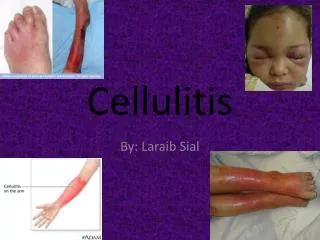
Tinea Pedis and Cellulitis of the Lower Extremities Alan L. Bisno, MD, FACP University of Miami School of Medicine
Factors Predisposing to Lower Extremity Cellulitis • Cutaneous disruption • Trauma, surgery • Burns • Ulcers • Varicella • Dermatophytes • Lymphedema, venous insufficiency • Obesity • Ischemia • IV drug abuse • Immunosuppression
Microbial Etiology of Eysipelas and Cellulitis • Classic erysipelas is virtually always due to beta-hemolytic streptococci (BHS) • The terms cellulitis and erysipelas are often used interchangeably in European literature. • Cellulitis can be due to many microorganisms. Most cases are due to BHS or Staphylococcus aureus. • Typical lower extremity cellulitis with diffuse spreading erythema is usually due to BHS of groups A, B,C, and G Baddour LM, BisnoAL. Amer J Med 1985; 79:155-159
Recurrent Cellulitis • Many patients with an episode of cellulitis experience repeated attacks. The percentage of patients in whom this occurs is variable, depending upon risk factors • In one study, 77% of such patients had abnormalities of lymphatic drainage on scintigraphy • It is believed that lymphatic damage is progressive with recurrent episodes, thus exacerbating the problem De Godoy JM et al. Lymphology 2000; 33: 177-180
Antimicrobial Prophylaxis of Recurrent Cellulitis • 31 patients with definite or presumptive streptococcal cellulitis of the LEs treated with 1.2 million units of benzathine penicillin G monthly for 12 months • 70 patients who declined and 14 who received incomplete prophylaxis served as controls • Recurrences rate was 12.9% in treated patients vs 19% in controls (NS) • BPG reduced recurrences in pts. without but not in those with predisposing factors Wang J-H et al. CID 1997; 25: 685-689
Antimicrobial Prophylaxis of Recurrent Cellulitis • 40 pts. with venous insufficiency or “lymphatic congestion” who had suffered ≥2 episodes of erysipelas during the previous 3 yrs. • 20 pts. received phenoxymethyl penicillin, 1 to 2 gm bid (or erythomycin 0.25 to 0.5 gm bid if allergic) for a median of 15 mos. with 2 recurrences vs 8 in 20 untreated controls (P=0.06) • Conclusion: continuous prophylaxis is indicated only in patients with a high recurrence rate. Sjoblom AC et al. Infection 1993; 21: 390-393
Tinea Pedis and Lower Extremity Cellulitis: Possible Pathophysiologic Mechanisms • The recovery of fungi decreases as athlete’s foot becomes progressively more severe. • Aerobic microflora expands as the disease becomes more and more serious. • Athlete’s foot represents a continuum from a relatively asymptomatic, scaling fungal eruption to a symptomatic, macerated, hyperkeratotic process that results from an overgrowth of resident organisms if the stratum corneum is damaged by preexistent fungi. • Overgrowth of the same organisms in normal, fungus-free interspaces does not produce lesions. Layden JJ, Kligman AM. Arch Dermatol1978; 114: 1466-1472
Tinea Pedis and Lower Extremity Cellulitis: Possible Pathophysiologic Mechanisms • With progression of the process of dermatophytosis, the normal protective skin barrier becomes macerated and friable. • As the process continues, the skin becomes debilitated and weakens as an effective barrier against infection. • Fissures may occur, providing a portal of entry for any opportunistic organism in the area, resulting in cellulitis. Day MR et al. Clinics in Podiatr Med Surg. 1996; 13; 759-766
Tinea Pedis and Lower Extremity Cellulitis • Initial studies focused upon patients who had undergone saphenous venectomy for coronary bypass grafts and subsequently experienced recurrent attacks of cellulitis due to BHS • Many of the infections were due to non-group A streptococci Baddour LM, Bisno AL. Ann Intern Med 1982; 97: 493-496; Greenberg J et al. Ann Intern Med 1982; 97: 565-566; Baddour LM, Bisno AL. Amer J Med 1985; 79: 155-159.
Tinea Pedis and Lower Extremity Cellulitis • Greenberg et al described 9 men aged 48 to 72 years who developed cellulitis in the saphenous venectomy extremity. • 5 of the 9 patients had recurrent episodes. • First infection was within 8 mos. post-op in 8 patients but one was 8.5 years later. Pos. blood cultures in 1 pt. yielded BHS. • All 9 had mild to severe tinea pedis of the involved leg. • No recurrences after aggressive topical or oral antifungal therapy. Greenberg J et al. Ann Intern Med 1982
Tinea Pedis and Lower Extremity Cellulitis • A subsequent report detailed 9 patients with post-CABG cellulitis, 5 of whom experienced from 2 to >20 recurrences. • 7 of these patients had tinea pedis, and in 2 instances, control of dermatophytosis was associated with cessation of attacks. Baddour LM, Bisno AL. JAMA 1984; 251: 1049-1052
Tinea Pedis and Lower Extremity Cellulitis • Semel and Goldin studied 20 patients with LE cellulitis. Patients with trauma, peripheral vascular disease or chronic leg ulcers were excluded. • Athlete’s foot was present in 20 (84%) of the 24 episodes studied. • BHS were isolated from ipsilateral toe web spaces in 17 (85%) of the 20 cases. • No BHS were recovered in 30 controls seen in a dermatologist’s office for treatment of Athlete’s foot but without cellulitis. (P<0.01) • Only BHS were recovered more frequently from patients than controls. Semel JD, Goldin H. Clin Infect Dis 1996; 23: 1162-1164.
Tinea Pedis and Lower Extremity Cellulitis • 30 Venezuelan patients with erysipelas but in otherwise good health • 13/30 (43%) had tinea pedis • In 7/30 (23%) tinea pedis was found to be the unique predisposing factor • No controls Roldan YB et al. Mycoses. 2000; 43; 181-183.
Tinea Pedis and Lower Extremity Cellulitis • Prospective but uncontrolled study of 62 hospitalized adults with cellulitis • Possible predisposing factors: • Dry skin—68% • Diabetes mellitus—50% • History of cellulitis—48% • Peripheral vasc. Dis.—40% • Trauma—32% • Tinea pedis—32% • S/P CABG—27% Koutkia P et al. Diag Microbiol Infect Dis 1999; 34: 325-327
Tinea Pedis and Lower Extremity Cellulitis • Case-control study of 167 patients hospitalized in 7 French hospitals for LE erysipelas and 249 hospitalized controls. • In multivariate analysis, significant risk factors were: disruption of cutaneous barrier (ulcer, wound, dermatosis, etc.), lymphedema, venous insufficiency, leg edema, overweight. • Toe-web intertrigo was present in 66% of patients and 23% of controls (OR, 6.6, 4.2 to 10.5) • Toe-web intertrigo was a strong risk factor (13.9, 7.2 to 27) with a population attributable risk of 61%. Dupuy A et al. BMJ 1999; 318: 1591-1594
FDA Adverse Event Reports of Cellulitis in Patients with Tinea Pedis • The majority of 13 these reports are more suggestive of allergy or hypersensitivity to the product rather than a true cellulitis.
Unresolved Issues • What is the risk of normal individuals with tinea pedis developing cellulitis at some time in their life? • Does this magnitude of risk justify a warning? • Are the BHS strains recovered from between the toes of patients with T. pedis and cellulitis truly the organisms responsible for cellulitis? I am unaware of reports of the same strain being recovered from toe cultures and cellulitis during a single attack of cellulitis.
Issues for the Committee • Tinea pedis is only one of a number of risk factors for the development LE cellulitis, but it is one of the most modifiable of such factors. • The committee might wish to add a caution about the importance of eradication of tinea pedis in patients wth such risk factors as lympedema, venous insufficiency, edema of the legs, marked obesity, saphenous venetctomy for CABG, or previous episodes of cellulitis.