Download
1 / 24
280 likes | 765 Views
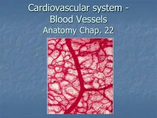
Cardiovascular system - Blood Vessels Anatomy Chap. 22. Arteries. Veins. Arterioles. Venules. Capillaries. Basic Anatomy of Circulatory routes. Carry blood towards the heart. Carry blood away from the heart. Connect capillaries to veins.

E N D
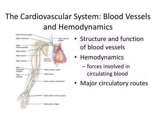
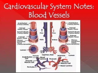
Arteries Veins Arterioles Venules Capillaries Basic Anatomy of Circulatory routes Carry blood towards the heart Carry blood away from the heart Connect capillaries to veins Control blood flow into capillaries & help regulate BP Allow for “exchange” (filtration/reabsorption) of O2/CO2, nutrients/wastes
Arteries & Veins Both are comprised of 3 layers of tissue surrounding “lumen” through which blood will flow: tunica interna, tunica media & tunica externa Structural difference between arteries & veins primarily due to differences in pressure of blood flowing within
Arteries & Veins • Tunica Interna – innermost endothelium of simple squamous epithelium + basement membrane • Arteries – have an “internal elastic lamina” of elastic CT to allow for expansion under pressure • Veins – may have “valves” (folds of endothelium + CT) to prevent backflow of blood due to low pressure
Arteries & Veins • Tunica Media – middle layer containing smooth muscle (for contractility/vasoconstriction) & elastic CT (for elasticity) • Arteries – have relatively thick tunica media allowing for significant vasoconstriction & elasticity • Elastic/conducting arteries – relatively more elastic tissue than smooth muscle; ie. aorta, pulmonary trunk, etc. • Muscular/distributing arteries - relatively more muscle tissue than elastic tissue; ie. brachial, femoral, etc. • Veins – relatively thin tunica media therefore no significant constriction/elasticity
Arteries & Veins • Tunica Externa (a.k.a. adventitia) – made of collagenous CT • Arteries – thin layer • Veins – thickest layer of vein, trying to support against gravity & low pressure
Arterioles & Venules • Very small, almost microscopic vessels with only 2 layers of tissue surrounding lumen • Arterioles – endothelium (tunica interna) + very thin layer of smooth muscle cells (tunica media); regulate blood flow to tissues & affect arterial blood pressure • Venules – endothelium (tunica interna) + thin layer of CT (tunica externa)
Capillaries • Microscopic, very thin-walled vessels comprised of endothelium with basement membrane; allows for filtration and reabsorption • Found in all tissues of the body except for those that are “avascular” • Usually form branching networks (“capillary beds”) within tissues for increased surface area • blood flow into capillaries may be regulated by “pre- capillary sphincters” • may have a central or “thoroughfare” channel that provides direct connection between “metarteriole” (terminal end of arteriole) & venule Capillaries can be classified as continuous, fenestrated, or sinusoids
Left common carotid artery Brachiocephalic trunk Left subclavian artery Ascending aorta (gives off coronary arteries) Aortic arch Thoracic (descending) aorta Abdominal aorta Common iliac arteries Circulatory Routes – Systemic circuit • Arterial blood from left ventricle into ascending aorta • Venous return to right atrium through SVC, IVC & coronary sinus
Cerebral circulation Cerebral arterial circle (“circle of Willis): Basilar artery (from union of vertebral arteries) + Internal carotid arteries
Hepatic portal circulation AORTA Venous blood flow from GIT & spleen to liver – ensures delivery of nutrients to liver first Hepatic artery Cystic vein Lt. gastric vein Splenic vein IVC LIVER Hepatic Veins (blood mixes in sinusioids) Hepatic Portal Vein Superior mesenteric vein Inferior mesenteric vein
Hepatic portal circulation • Cystic vein, left gastric vein, splenic vein, inferior & superior mesenteric veins Hepatic portal vein • Hepatic portal vein (deoxygenated/nutrient rich blood) + Hepatic artery (oxygenated blood) sinusoids of liver • Sinusoids of liver Hepatic veins IVC
Fetal Circulation Placenta – O2/CO2 & nutrient/waste between mom & baby Umbilical cord – (2)umbilical arteries (baby mom) & (1)umbilical vein (mom baby) Umbilical vein (O2/nutrient rich blood) hepatic portal vein & ductus venosus IVC Rt. atrium Rt. Atrium some blood to rt. Ventricle, most shunts across foramen ovale in interatrial septum lt. atrium lt. ventricle
Fetal Circulation Blood from Rt. Ventricle pulmonary trunk across ductus arteriosus to aorta Aorta systemic arteries internal iliac arteries umbilical arteries placenta
Fetal Circulation- Changes at Birth • Umbilical vein ligamentum teres (round ligament) • Umbilical arteries lateral umbilical ligaments • Ductus venosus ligamentum venosum • Foramen ovale fossa ovalis • Ductus arteriosus ligamentum arteriosum • Placenta delivered (“afterbirth”)