Download

1 / 15
340 likes | 2.27k Views
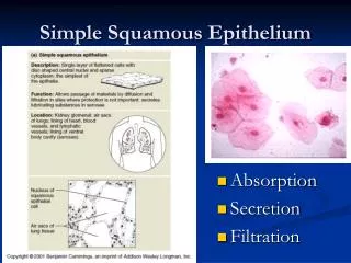
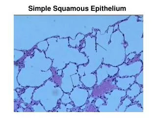
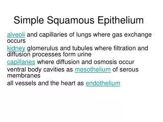
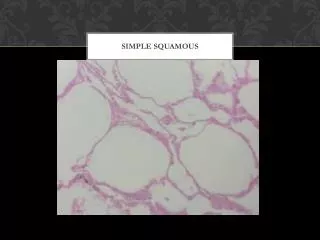
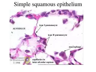
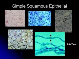
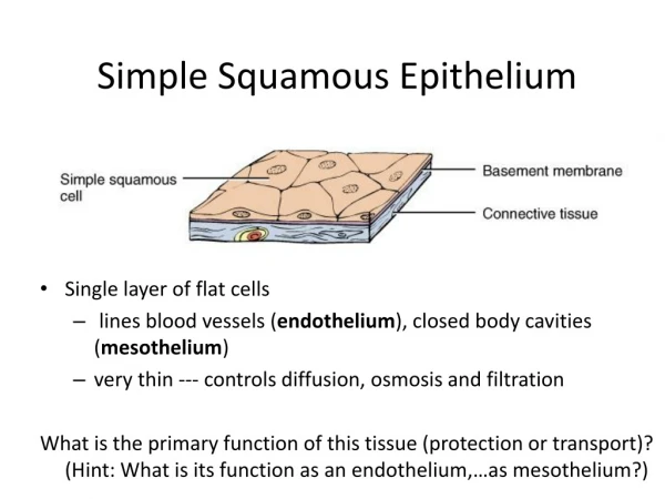
Simple Squamous Epithelium. Single layer of flat cells lines blood vessels ( endothelium ), closed body cavities ( mesothelium ) very thin --- controls diffusion, osmosis and filtration

E N D
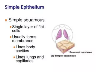
Simple Squamous Epithelium • Single layer of flat cells • lines blood vessels (endothelium), closed body cavities (mesothelium) • very thin --- controls diffusion, osmosis and filtration What is the primary function of this tissue (protection or transport)? (Hint: What is its function as an endothelium,…as mesothelium?)
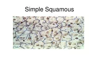
Section of intestine showing serosa Surface view of lining of peritoneal cavity Examples of Simple Squamous Epithelium Is this endothelium, mesothelium, or neither?
Simple Cuboidal Epithelium • Single layer of cube-shaped cells viewed from the side Where would you find simple cuboidal epithelium in the body? Does it typically function in protection or transport in these locations?
Example of Simple Cuboidal Epithelium • Sectional view of kidney tubules Why do some cells look as if they do not have a nucleus?
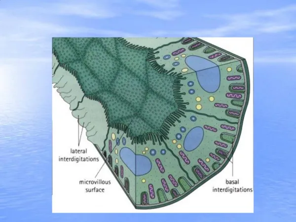
Nonciliated Simple Columnar • Unicellular glands =goblet cells secrete mucus • lubricate GI, respiratory, reproductive and urinary systems • Microvilli = fingerlike projections of cell membrane Describe the differences between cilia and microvilli in terms of both their structure and function. Are you likely to find them on the same tissue? Why or Why not?
Example: Nonciliated Simple Columnar Epithelium • Section from small intestine The term “brush border” is often used to describe this tissue. What does it refer to?
Ciliated Simple Columnar Epithelium • Single layer rectangular cells with cilia • Mucus from goblet cells moved along by cilia
Example Ciliated Simple Columnar Epithelium • Section of uterine tube What is another name for this organ?
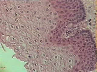
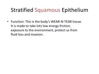
Stratified Squamous Epithelium • Several cell layers thick • Surface cells flat • Keratinized = surface cells dead and filled with keratin • Example…? • Nonkeratinized = no keratin in moist, living cells at apical surface • Example …? What is the primary function of this epithelium? If this epithelium is a portion of a mucus membrane, where does the mucus come from?
Example of Stratified Squamous • Section of vagina
Stratified Cuboidal Epithelium • Multilayered • Surface cells cuboidal • rare (only found in sweat gland ducts & male urethra)
Stratified Columnar Epithelium • Multilayered • Surface cells columnar • Rare (very large ducts & part of male urethra)
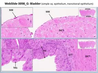
Multilayered Surface cells varying in shape from round to flat if stretched Lines hollow organs of the urinary tract that expand from within Transitional Epithelium List the expandable organs of the urinary tract.
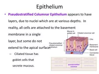
Pseudostratified Columnar Single cell layer All cells attach to basement membrane but not all reach free surface Nuclei at varying depths Name one location where you would find this epithelium. How would you describe its function?