Download
1 / 28
340 likes | 1.08k Views
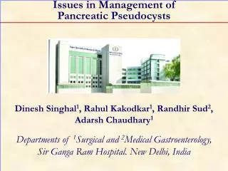
Issues in Management of Pancreatic Pseudocysts Dinesh Singhal 1 , Rahul Kakodkar 1 , Randhir Sud 2 , Adarsh Chaudhary 1 Departments of 1 Surgical and 2 Medical Gastroenterology, Sir Ganga Ram Hospital. New Delhi, India Abstract

E N D
Issues in Management ofPancreatic Pseudocysts Dinesh Singhal1, Rahul Kakodkar1, Randhir Sud2, Adarsh Chaudhary1 Departments of 1Surgical and 2Medical Gastroenterology,Sir Ganga Ram Hospital. New Delhi, India
Abstract Pancreatic pseudocysts (PPs) comprise more than 80 % of the cystic lesions of the pancreas and cause complications in 7-25% of patients with pancreatitis or pancreatic trauma. The first step in the management of PPs is to exclude a cystic tumor. A history of pancreatitis, no septation, solid components or mural calcification on CT scan and high amylase content at aspiration favor a diagnosis of PP. Endoscopic ultrasound (EUS)-guided FNAC is a valuable diagnostic aid. Intervention is indicated for PPs which are symptomatic, in a phase of growth, complicated (infected, hemorrhage, biliary or bowel obstruction) or in those occurring together with chronic pancreatitis and when malignancy cannot be unequivocally excluded. The current options include percutaneous catheter drainage, endoscopy and surgery. The choice depends on the mode of presentation, the cystic morphology and available technical expertise. Percutaneous catheter drainage is recommended as a temporizing measure in poor surgical candidates with immature, complicated or infected PPs. The limitations include secondary infection and pancreatic fistula in 10-20% of patients which increase complications following eventual definitive surgery. Endoscopic therapy for PPs including cystic-enteric drainage (and transpapillary drainage), is an option for PPs which bulge into the enteric lumen which have a wall thickness of less than 1 cm and the absence of major vascular structures on EUS in the proposed tract or those which communicate with the pancreatic duct above a stricture. Surgical internal drainage remains the gold standard and is the procedure of choice for cysts which are symptomatic or complicated or those having a mature wall,. Being more versatile, a cystojejunostomy is preferred for giant pseudocysts (>15cm) which are predominantly inframesocolic or are in an unusual location. In PPs with coexisting chronic pancreatitis and a dilated pancreatic duct, duct drainage procedures (such as longitudinal pancreaticojejunostomy) should be preferred to a cyst drainage procedure.
Issues • Observation or intervention? • Pseudocyst or tumor ? • Management strategy • Endoscopy or surgery ? • Complicated pseudocyst ?
Indications for Intervention • Absolute indications • Symptomatic • Chronic pseudocysts* • In a phase of growth • Complications • Malignancy ?
Indications for Intervention • Relative indications • Duration: more than 6 weeks • Size: greater than 6 cm • Pancreatic duct abnormalities(stricture, stone, rupture) • Multiple cysts*
Expectant Management • Asymptomatic • Uncomplicated • Stable or decreasing size
Beware of a Cystic Tumor ! Cystic tumor erroneously drained by ‘cystogastrostomy’ October 2002 December 2003 Enhancing walls, solidcontent, evidence of neoplasm Cystic tumour misinterpreted as pseudocyst
Previous pancreatitis/trauma Imaging (CT, US): Single, non-loculated No septae or solid components Thin wall (<4mm) MRCP/ERCP No history of pancreatitis Imaging: Often multilocular Septae or solid components+ Thick walled MRCP/EUS ± FNA /ERCP Pseudocyst vs. Cystic Tumor Duct-cyst connectionin 65% Noduct-cyst connection
Pseudocyst vs. Cystic Tumor Pseudocyst Cystic tumor
Cyst Fluid Analysis [1] Lewandrowski KB, et al. Ann Surg 1993, 217:41-7.[2] Brugge WR, et al. N Engl J Med 2004, 351:1218-26.
Pseudocyst vs. Cystic Tumor • Retrospective study of 21 cystic neoplasms; 8 diagnosed pseudocysts • Only one patient had a history of pancreatitis • 7/8 CT scans lacked features which were suspicious of neoplasm • 16/18 investigations ( ERCP, cyst fluid analysis, angiography) unhelpful • A mucinous cystic neoplasm is more likely to be misdiagnosed as a pseudocyst • 5/13 MCA misdiagnosed; 2 underwent cystenterostomy • At imaging, classical findings of neoplasia (septae, wall calcification and papillary projections) were absent in 38% of cases [3] Martin I, et al. Br J Surg 1998; 85:1484-6. [4] Scott J, et al. Clin Radiol 2000, 55:187-92.
Pseudocyst vs. Cystic Tumor No imaging is infallible ! It is better to resect a pseudocystthan to drain a tumor !
Uncomplicated Cyst • Bulge into stomach/duodenum* • No solid tissue/vessels (EUS)* • Wall thickness 0.5-1cm (EUS) • Technical expertise available Pseudocyst entered Endoscopicdrainage Tract dilated Drain placed [5] Kahaleh M, et al. Endoscopy 2006; 38:355-9. [6] Sanchez Cortes E, et al. Gastrointest Endosc 2002; 56:380-6. [7] Sriram PV, et al. Endoscopy 2005; 37:231-5.
Surgical Strategy (I) • Symptomatic maturepseudocyst with bulging intothe posterior gastric wall Pseudocyst with mature wallbulging into the stomach Anterior Gastric Wall Pseudocyst openedthrough posteriorgastric wall Cystogastrostomy* Cystogastrostomy-intraoperative
Surgical Strategy (II) • Symptomatic maturepseudocyst with infracolicbulging or giant pseudocyst Giant pseudocyst Colon Cystojejunostomy Pseudocyst Large pseudocystin infracolic position
Surgical Strategy (III) • Symptomatic mature pseudocyst + dilated main pancreatic duct Pseudocyst withdilated mainpancreatic duct Partington-Rochelle
Surgical Strategy (IV) • Symptomatic maturemultiple pseudocystsin unusual locations+ dilated mainpancreatic duct Medistinal pseudocyst Partington-Rochelle Duct-cyst communication (forcep)
Complications • Sepsis • Biliary obstruction • Hemorrhage • Sinistral portal hypertension • Duodenal obstruction
Sepsis • Infected pseudocyst (abscess)not amenable toimage-guided drainage Intracystic air Infected pseudocyst ‘Abscess’ External drainage Pseudocyst (Risk of pancreatic fistula morbidity:10-15%) Drained externally [8] Adams DB, Anderson MC. Ann Surg 1992; 215:571-8. [9] Heider R, et al. Ann Surg 1999; 229:781-7.
Biliary Obstruction (I) • Complicated pseudocystwith immature wall Pseudocyst with immature walls External drainage (Risk of pancreatic fistula morbidity:10-15%) Drained percutaneouslythrough safe infracolic window
Biliary Obstruction (II) • Mature pseudocyst withbiliary obstruction • Not amenable to ERC Mature pseudocyst abuttingstomach and causingbiliary obstruction Internal drainage (Surgical/Endoscopic) Obstruction relievedafter cystogastrostomy
Recent hemorrhage Pseudoaneurysm Hemorrhage (I) • Intracystic bleeding withmature wall (failedangioembolization) Pseudocyst with bleed Ligation/Packing
Pseudoaneurysm Hemorrhage (II) • Pseudoaneurysmwithout bleeding Pseudoaneurysm of splenic artery Angioembolization/Resection
Sinistral Portal Hypertension • Sinistral portal hypertensionwith fundal variceal bleeding,dilated main pancreatic duct,and pancreatic pain Dilated MPD Collaterals in splenic hilum Splenectomy +Ductal drainage Small pseudocyst
Duodenal Obstruction Pseudocyst causing extrinsiccompression of the duodenum • Duodenal obstruction Duodenal obstruction Gastrojejunostomy*
Key Points • Rule out cystic tumor • Endoscopic drainage in selected patients • Surgery - gold standard for pseudocysts: • Giant • Complicated • Associated with ductal abnormalities
Keywords Cystadenoma; Cysts; Endosonography; Pancreas; Pancreatic Pseudocyst; Surgery Abbreviations MCA: mucinous cystadenoma; MCAC: mucinous cystadenocarcinoma; SCA: serous cystadenoma CorrespondenceAdarsh ChaudharyDepartment of Surgical GastroenterologySir Ganga Ram HospitalNew DelhiIndia 110060Phone: +91-11.4225.2226Fax: +91-11.4225.2224E-mail: adarsh@nda.vsnl.net.in
References • Lewandrowski KB, et al. Ann Surg 1993, 217:41-7. [Full text] • Brugge WR, et al. N Engl J Med 2004, 351:1218-26. [Full text] • Martin I, et al. Br J Surg 1998; 85:1484-6. [Full text] • Scott J, et al. Clin Radiol 2000, 55:187-92. [Full text] • Kahaleh M, et al. Endoscopy 2006; 38:355-9. [Full text] • Sanchez Cortes E, et al. Gastrointest Endosc 2002; 56:380-6. [Full text] • Sriram PV et al. Endoscopy 2005; 37:231-5. [Full text] • Adams DB, Anderson MC. Ann Surg 1992; 215:571-8. [Full text] • Heider R, et al. Ann Surg 1999; 229:781-7. [Full text]