Download
1 / 3
30 likes | 37 Views
The current imaging methods for the prostate cancer diagnosis<br>are complicated and partially invasive. Therefore, a key challenge<br>for prostate cancer detection is to use a simple and non-invasive<br>method. Imaging plays a crucial role in the non-invasive identification,<br>localization and grading of prostate carcinoma. In this paper, we<br>demonstrate the possibility using of polarized a near infrared (NIR)<br>light for the in vitro prostate cancer detection and imaging. Because<br>the intensity of NIR light passing through the cancerous outgrowth<br>is lower than the intensity of NIR light passing through the noncancerous one, the cancerous formations are differentiated as the<br>dark areas in the relatively white background. Specially developed<br>software analyses and processes a distribution of intensities of<br>the grayscale images, measures the ratios of their strength, and<br>determines the rate of prostate malignancy. Obtained results may<br>hold promise to make an important contribution to the diagnosis of<br>prostate cancer in the early stage of its development

E N D
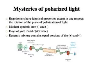
Partsvania et al., J Clin Exp Oncol 2017, 7:1 DOI: 10.4172/2324-9110.1000208 Journal of Clinical & Experimental Oncology Research Article a SciTechnol journal [8,9]. On the other hand it is well known, that polarized light has ability to eliminate details which are not noticeable in ordinary light. Thereby in this paper, we demonstrate that polarized NIR light can be used successfully for the visualization of cancerous outgrowth in the prostate, obtained by the radical prostatectomy. Methods Experimental materials, such as prostate glands, were obtained from the radical prostatectomy. Light emitted diodes (LED) s were utilized as the light sources (QT Brightek Company, USA), emitting an infrared light in the 850-900 nm range of the optical spectrum. The consuming power of LEDs was low, in the range of 0, 08-0, 14 Watt and therefore, they did not cause any heating and damaging of the prostate tissues. To observe the prostate glands in the NIR spectrum, a CCD camera (IR 1000, Dage-MTI, USA) connected to a personal computer was used. To avoid a possible artifact caused by the visible light, experiments were carried out in the darkness. Every form of light represents electromagnetic waves, whose electric field vectors vibrate in all planes that are perpendicular with respect to the direction of propagation. This is true as to visible wavelengths of spectrum as to invisible infrared waves. If the electric field vectors are restricted to a single plane by filtration of the beam with specialized materials, called polarizers, then the light is referred to as plane or linearly polarized with respect to the direction of propagation. Wave passing through the polarizer is subsequently blocked by the second polarizer, if this polarizer is oriented horizontally with respect to the electric field vector in the light wave. If optical isomer is placed on the path of the polarized beam the E vector polarization angle can be shifted. Consequently, corresponding rotation of second polarizer causes passing of the light through it. For polarizing of Infrared (IR) light we used a Linear Polarizer APIR29- 020 of the American Polarizers, Inc. In experiments LED was placed outside the prostate gland, from the bottom side, enabling NIR light to totally penetrate a polarize film then prostate tissue and another polarize film. In the Figure 1 is shown the experimental setup: Investigation of the prostate glands in the polarized infrared light was performed immediately after radical prostatectomy. Afterward histo-morphological examination was executed based on the standard methods [10] that enable a tumor to be located precisely in the prostate tissue. Because the investigations were carried out on the base of a standard method, we did not consider including in our paper an illustrative stuff of the histo-morphological examinations. Accordingly, a location of each cancerous outgrowth was determined using two methods: one which was detected by histo- morphological examination and other, by means of the polarized NIR investigations. To compare the transillumination images of non-cancerous tissues with the cancerous ones, we have elaborated PC software. That separately calculates the average values of the brightness for the potentially healthy tissue and for the malignant one in an IR image and calculates their ratio. Software calculates a 95% confidence interval. In our case, after the examination of 64 cancerous prostates we obtained Utilization of the Polarized Infrared Light for Prostate Cancer Visualization in Isolated Prostates Besarion Partsvania1, Giorgi Kochiashvili2* and Alexander Khuskivadze2 Abstract The current imaging methods for the prostate cancer diagnosis are complicated and partially invasive. Therefore, a key challenge for prostate cancer detection is to use a simple and non-invasive method. Imaging plays a crucial role in the non-invasive identification, localization and grading of prostate carcinoma. In this paper, we demonstrate the possibility using of polarized a near infrared (NIR) light for the in vitro prostate cancer detection and imaging. Because the intensity of NIR light passing through the cancerous outgrowth is lower than the intensity of NIR light passing through the non- cancerous one, the cancerous formations are differentiated as the dark areas in the relatively white background. Specially developed software analyses and processes a distribution of intensities of the grayscale images, measures the ratios of their strength, and determines the rate of prostate malignancy. Obtained results may hold promise to make an important contribution to the diagnosis of prostate cancer in the early stage of its development. Keywords Prostate cancer; Imaging; Polarized infrared light Introduction Despite significant advances achieved in prostate cancer diagnosis it remains the second cause of cancer death in men worldwide [1]. The reason is that existent methods of diagnosis are not effective to detect cancer at early stage of development. Besides imaging method such are magnetic resonance imaging (MRI) [2,3] and Positron-emission tomography (PET) [4,5] are partially invasive. These methods are complicated and expensive and could not be used in ordinary clinics. These limitations of mentioned imaging methods of detection prostate cancer have led to seek of alternative techniques. The use of near infrared (NIR) optical methods for diagnosing and detecting cancer has generated considerable interest [6,7]. Optical methods are advantageous, because they are non-invasive, fast, relatively inexpensive, pose no risk of ionizing radiation, and in addition, NIR light can easily penetrate thick tissues. In our previous works we showed possibility usage of NIR transillumination technology for prostate cancer detection in vitro *Corresponding author: Giorgi Kochiashvili, Tbilisi State Medical University, Tbilisi, 0160, Georgia, Tel: (995) 599505563; E-mail: kochiashviligiorgi@hotmail.com Received:November 06,2017Accepted:November 22, 2017Published: November 30, 2017 All articles published in Journal of Clinical & Experimental Oncology are the property of SciTechnol, and is protected by copyright laws. Copyright © 2017, SciTechnol, All Rights Reserved. International Publisher of Science, Technology and Medicine
Citation: Partsvania B, Kochiashvili G, Khuskivadze A (2017) Utilization of the Polarized Infrared Light for Prostate Cancer Visualization in Isolated Prostates. J Clin Exp Oncol 7:1. doi: 10.4172/2324-9110.1000208 interval of above mentioned ratios within 0.169417-0.239595 range. Thereafter, for new unknown prostate NIR transillumination image will be analysed and software will calculate above mentioned ratio. It will compare a meaning of this calculation with that confidence interval and detects with 95% probability cancerous malignancy of this prostate. Results and Discussion In this investigation, we have carried out a series of experiments in which were examined 64 cancerous and 30 non-cancerous prostate glands. The experiment revealed that prostate tissue is able to shift polarization plane of IR irradiation. The effect of shifting of E vector direction does not depend on existence of cancer; rather it is a feature of prostate tissue. The infrared transillumination images of the non- cancerous prostates in polarized light are characterized by a nearly homogeneous surface, in which a distribution of light intensities are equal everywhere (Figure 2). IR images of cancerous prostate in non polarized and polarised lights are shown in the Figure 3. Comparison of IR images obtained with nonpolrized and polarized irradiations revealed that light polarization gives opportunity to detect cancerous outgrowths in the areas which were not notable in the nonpolrized light. The differences in the optical properties between the cancerous and non-cancerous cells can be caused due to the following reason: as it is known generally, the cancerous cells are characterized by non-controlled and chaotic division. Normal cells have one nucleus and one nucleolus. Chromatins are threadlike before the division. Cancerous cells have more than one large irregular shaped nucleus and nucleolus and condensed chromatins. Due to this reason a light penetration in normal and in cancerous cells is not the same, healthy cells are nearly transparent to light, whereas cancerous cells are much less transparent to light [11]. Moreover, this decision is fair with respect to the NIR spectrum window, in which the biological tissues show very low absorption and auto-fluorescence. Conclusion In summary, it has been shown that the examination of the penetration of NIR light through the prostate tissue is a promising imaging modality in the field of prostate cancer targeting and visualization in the early stage of development. Our experiments have shown that a passage of NIR light through the cancerous and non-cancerous prostate tissues significantly differs from each other. Therefore, the infrared images of cancerous outgrowth are observed as the dark areas in the bright background. It is shown that polarized NIR significantly increases possibility to detect cancerous outgrowths. The minimized photo-damage, low auto-fluorescence, high penetration (a) (b) Figure :1 a) LED and CCD camera are shown with yellow arrows. Polarizers are shown with black arrows. b) IR light passes through polarizer 1 (indicated by the yellow arrow), then the prostate tissue and a second polarizer (indicated by the black arrow) and enters into the CCD camera. Figure 2: IR image of a non-cancerous prostate in polarized IR light. Diagnosis was benign hyperplasia. • Page 2 of 3 • Volume 7 • Issue 1 • 1000208
Citation: Partsvania B, Kochiashvili G, Khuskivadze A (2017) Utilization of the Polarized Infrared Light for Prostate Cancer Visualization in Isolated Prostates. J Clin Exp Oncol 7:1. doi: 10.4172/2324-9110.1000208 a b Figure 3: IR images of cerous prostate in non polarized and polarised lights. a) IR image of prostate in nonpolarizedl light. Cancerous outgrowths are indicaded with arrows. b) IR images of the same prostate in polarized IR light. Structures indicated with yellow arows are not noticable in the left figure (a) corresponding to non polarized IR ligth. Afterward histo-morphological investigations indicated, that these strucutres were cancerous outgrowths. depth and NIR LED based simple technique offers unique advantages for the visualization and diagnosis of prostate malignancy. However, it should be noted that the further studies are needed in order to identify and differentiate the infrared images of cancerous outgrowth with the other possible symptoms, such as inflammation. 5. Mauer T, Beer AJ, Souvatzoglou M, Holzapfel K, Kubeler H, et al. (2014) 68Gallium-labelled ligand of prostate-specific membrane antigen (PSMA) for the evaluation of recurrent prostate cancer using PET/CT andPET/MR imageing. Eur Urol 13: 726. 6. Champaign JL, Cederbom GJ (2000) Advances in breast cancer detection with screening mammography.Ochsner J 2: 33-35. Acknowledgment 7. Tromberg BJ, Cerussi A, Shah N, Compton M, Durkin A, et al. (2005) Imaging in breast cancer: Diffuse optics in breast cancer: Detecting tumors in pre- menopausal women and monitoring neoadjuvant chemotherapy. Breast Cancer Res 7: 279 -285. This work was supported by ISTC foundation, project #G-2188. References 8. Khuskivadze A, Kochiashvili D, Partsvania B (2013) Near infrared radiation in diagnosis of prostate cancer: Preliminary results. Urology82: S1-S334. 1. Siegel RL, Miller KD, Jemal A (2015) Cancer statistics, 2015. CA Cancer J Clin 65: 5-29. 9. Khuskivadze A, Partsvania B, Kochiashvili D (2014) Visualization of human prostate cancer using infrared radiation. Urology 84: S304. 2. Hendee WR, Morgan CJ (1984) Magnetic resonance imaging. Part I-Physical Principles. West J Med141: 491-500. 10. Svanholm H, Starklin H, Gundersen HJ, Fabricius J, Barlebo H, et al. (1989) Reproducibility of histomorphologic diagnoses with special reference to the kappa statistic. APMIS 97: 689-698. 3. Luechinger R, Duru F, Candinas R, Boesiger P (2004) Safety considerations for magnetic resonance imaging of pacemaker and ICD patients. Herzschrittmachertherapie und Elektrophysiologie 15: 73-81. 11. Ertefai S, Profio AE (1985) Spectral transmittance and contrast in breast diaphanography. Med Phys 12: 393-400. 4. Eiber M, Nekolla SG, Maurer T, Weirich G, Wester HJ, et al. (2014) (68) Ga-PSMA PET/MR with multimodality image analysis for primary prostate cancer. Abdom Imaging 40: 1769-1771. Author Affiliations Top 1Institute of Cybernetics, Georgian Technical University, Tbilisi, 0186, Georgia 2Tbilisi State Medical University, Tbilisi, 0160, Georgia Submit your next manuscript and get advantages of SciTechnol submissions 80 Journals 21 Day rapid review process 3000 Editorial team 5 Million readers More than 5000 Quality and quick review processing through Editorial Manager System Submit your next manuscript at ● www.scitechnol.com/submission • Page 3 of 3 • Volume 7 • Issue 1 • 1000208